1. Introduction to the Molecular Landscape
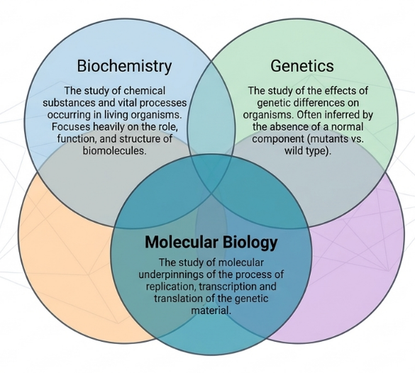
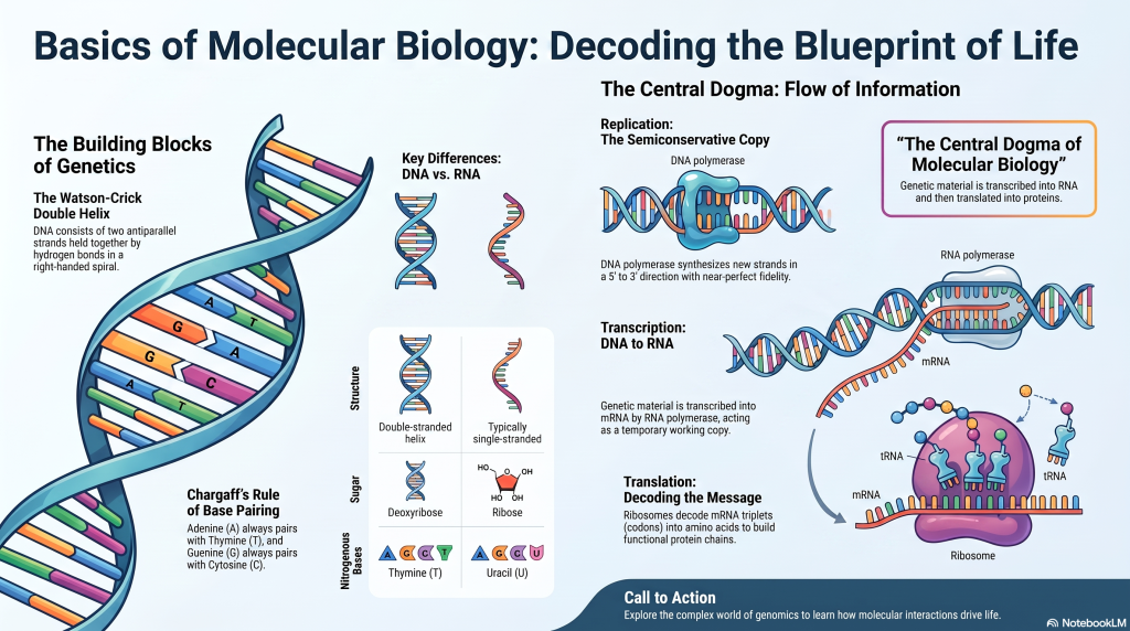
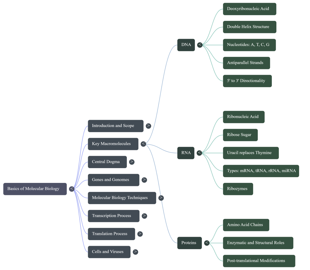
Molecular biology is the rigorous study of life at the molecular level, serving as the critical intersection where genetics and biochemistry converge to explain the phenomena of life through macromolecular properties. This discipline focuses primarily on characterizing the structure, function, and regulatory relationships between the two primary macromolecules: nucleic acids (the repository of genetic information) and proteins (the active agents of biological systems). Specifically, the field examines the intricate orchestration of DNA, RNA, and protein biosynthesis, as well as the regulatory mechanisms that govern these interactions.
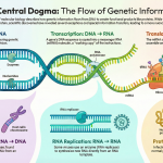
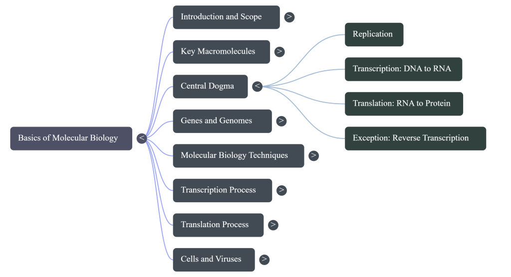
The framework for this field is traditionally defined by the “Central Dogma”—the flow of information from DNA to RNA and subsequently to proteins. However, this model is currently evolving to accommodate a more complex landscape, including the discovery of novel regulatory functions for non-coding RNA and reverse transcription. Molecular biology is defined by a lack of “definite lines” between it and other core disciplines. In contemporary research, molecular biology and biochemistry have become nearly interchangeable, with the former providing the underpinnings for replication and transcription, while biochemistry investigates the chemical substances and vital processes occurring in living organisms. Genetics provides the context of how these components—or their absence—manifest as phenotypic traits, while biophysics offers a “from the ground up” tradition of analyzing biomolecules through physical laws. This synergy creates a unified field essential for the modern understanding of cellular life and its historical milestones.
2. The Molecular Revolution: A Chronology of Discovery
A profound understanding of the history of molecular biology is essential for contextualizing modern genetic engineering. The field’s maturation in the 1930s and 1940s was driven by a strategic shift in research methodology: moving from the traditional genetic mainstay Drosophila to more technically appropriate model organisms, such as the fungus Neurospora and bacteriophages. This transition allowed researchers to investigate life at its most fundamental chemical level.
Key Milestones in Molecular Discovery
| Year | Researcher(s) | Breakthrough Contribution |
|---|---|---|
| 1860s | Gregor Mendel | Proposed “factors” (genes) as the basic units of trait inheritance in pea plants. |
| 1910 | Thomas Hunt Morgan | Demonstrated that genes reside at specific locations (loci) on chromosomes. |
| 1928 | Frederick Griffith | Showed that genetic information could be transferred between bacterial strains. |
| 1944 | Avery, MacLeod, and McCarty | Provided definitive proof that genes are composed of DNA. |
| 1952 | Hershey and Chase | Confirmed DNA as the genetic material using bacteriophage T2. |
| 1953 | Watson and Crick | Elucidated the double-helical structure of the DNA molecule. |
| 1984 | Kary Mullis | Invented the Polymerase Chain Reaction (PCR) for enzymatic DNA amplification. |
The transition toward quantitative research was catalyzed by George Beadle and Edward Tatum. Their “one gene, one enzyme” hypothesis—positing that mutations in specific genes cause errors in specific metabolic pathways—demonstrated a precise relationship between genetic material and biochemical activity. This shift from descriptive to mechanistic biology established the foundation for our modern comprehension of gene structure and function.
3. Genetic Architecture: DNA, RNA, and the Chemical Basis of Heredity
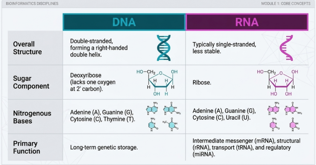
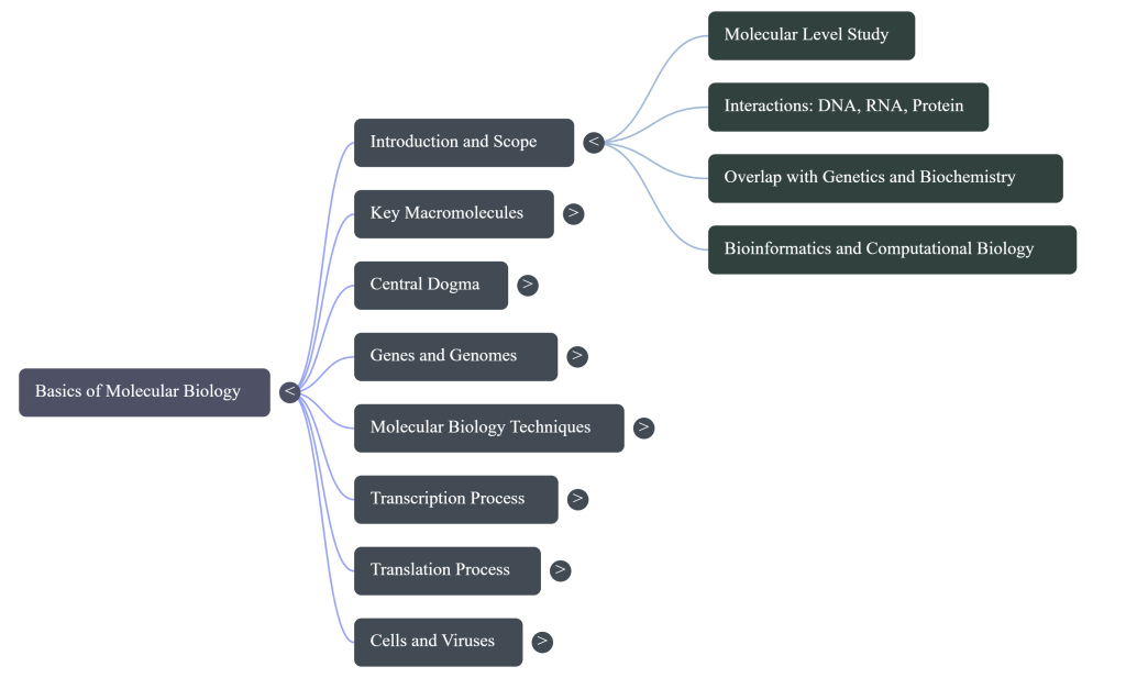
Nucleic acids are the primary informational macromolecules of life, structured to facilitate the storage, replication, and transmission of biological data. These polymers are composed of nucleotide subunits, each consisting of a phosphate group, a pentose sugar ring, and a nitrogenous base.
Biochemical Differentiators: DNA vs. RNA
The Watson-Crick Double-Helix Model
The DNA double helix consists of two complementary strands twisting in a right-handed spiral. These strands are oriented antiparallel to one another. To conceptualize this, imagine walking down the sugar-phosphate backbone: on one strand, you would encounter the 5′ carbon followed by the 3′ carbon in a head-to-tail fashion; on the opposing strand, you would encounter them in the reverse order (3′ then 5′). The structural integrity of this helix is maintained by specific hydrogen bonding: Adenine (A) pairs with Thymine (T) via two hydrogen bonds, while Guanine (G) pairs with Cytosine (C) via three hydrogen bonds.
The 5′-3′ directionality of DNA is the “So What?” layer of genetic architecture. Cellular enzymes are restricted to recognizing just one direction; consequently, all nucleic acid synthesis occurs strictly in the 5′-3′ direction. The 3′ hydroxyl group acts as a critical nucleophile in the dehydration reaction required to add new monomers. Without this specific chemical orientation, the high-fidelity replication and transcription required for life would be impossible.
4. DNA Replication
DNA replication is the fundamental biological process by which a cell makes an identical copy of its DNA, serving as the basis for biological inheritance. This process is described as semiconservative, meaning that each of the two strands of the original double-stranded DNA molecule serves as a template for the synthesis of a new, complementary partner strand. Consequently, the result of replication is two identical DNA molecules, each consisting of one original “parental” strand and one newly synthesized “daughter” strand.
The Mechanism of Replication
Replication is a highly complex and tightly regulated process that follows specific rules of molecular biology:
- Directionality: DNA synthesis occurs strictly in the 5′ to 3′ direction. This is because the enzyme responsible for synthesis, DNA polymerase, can only add new nucleotides to the exposed 3′ hydroxyl group of an existing chain.
- Complementary Base Pairing: The redundancy of information in the double helix allows for accurate copying; adenine (A) always pairs with thymine (T), and guanine (G) always pairs with cytosine (C).
- Energy Source: The energy required to form the chemical (phosphodiester) bonds between nucleotides comes from the removal of two phosphate groups from the incoming nucleoside triphosphates.
Key Enzymes and Proteins
A “replisome” of proteins works together at a structure called the replication fork:
- DNA Polymerase: The primary enzyme family that carries out replication by assembling new DNA strands. It cannot start a new strand from scratch and requires a short fragment called a primer (usually RNA) to begin.
- Helicase: This enzyme unwinds the double helix by breaking the hydrogen bonds between the base pairs.
- Topoisomerase: As DNA is unwound, it creates physical twists and tension further down the line; topoisomerases cut and swivel the DNA strands to relieve this stress.
- Primase: An enzyme that builds the necessary RNA primers to give DNA polymerase a starting point.
- Ligase: This enzyme acts as “molecular glue,” joining together fragments of DNA to create a continuous strand.
- Single-strand binding proteins: These bind to the separated single strands of DNA to prevent them from prematurely re-forming a double helix or folding into interfering structures.
Leading vs. Lagging Strands
Because the two strands of DNA are antiparallel (running in opposite directions) and DNA polymerase can only synthesize in one direction, the two new strands are produced differently:
- Leading Strand: This strand is synthesized continuously in the same direction that the replication fork is moving.
- Lagging Strand: This strand is synthesized discontinuously in the opposite direction of the fork movement. It is made in short segments known as Okazaki fragments, which are later joined together by DNA ligase.
Accuracy and Regulation
DNA replication is remarkably accurate, making fewer than one error for every 10 million nucleotides added. This high fidelity is maintained through proofreading mechanisms where the DNA polymerase can detect, remove, and correct mismatched bases.
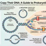
In eukaryotic cells, replication is strictly controlled within the S phase (synthesis phase) of the cell cycle. Unlike bacteria, which typically have a single “origin” of replication on a circular chromosome, eukaryotes initiate replication at multiple points along their long linear chromosomes. To protect the ends of these linear chromosomes from being lost or degraded during repeated rounds of replication, they are capped with repetitive sequences called telomeres.
5. Transcription
Transcription is the biological process of creating an equivalent RNA copy from a specific sequence of DNA. It serves as the first step of gene expression in the central dogma of molecular biology, where genetic information is transferred from DNA to RNA before being translated into proteins.
The Role of RNA Polymerase
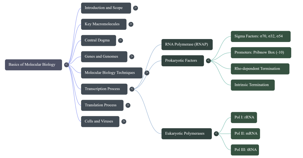
The primary enzyme responsible for this process is RNA polymerase (RNAP). Unlike DNA polymerase, RNA polymerase can initiate synthesis de novo and includes its own helicase activity to unwind the DNA double helix. It builds the RNA chain by adding ribonucleotides to the 3′ end of the transcript, meaning synthesis always occurs in the 5′-3′ direction.
The Process of Transcription
Transcription typically proceeds through three main stages:
- Initiation: RNA polymerase binds to specific DNA sequences called promoters, which signal where a gene begins. In bacteria, the polymerase must first bind to a specificity factor called a sigma (σ) factor to recognize these promoter regions, such as the Pribnow box. This stage transitions from a “closed complex” (where DNA is still wound) to an “open complex” as the DNA unwinds.
- Elongation: The enzyme moves along the DNA template, unwinding the helix and synthesizing a complementary RNA strand. In eukaryotes, these RNA chains can be remarkably long, such as the 2.4 million nucleosides of the dystrophin gene.
- Termination: The process ends when the polymerase encounters specific sequences known as terminators. In prokaryotes, this occurs via two well-known mechanisms: intrinsic termination, where the RNA itself forms a hairpin structure that dislodges the enzyme, or Rho-dependent termination, which requires a specific protein called the ρ (rho) factor.
Key Differences Between Organisms
The environment and regulation of transcription vary significantly between cell types:
- Prokaryotes: Transcription occurs in the cytoplasm. Because there is no nuclear membrane to separate the genetic material from the ribosomes, transcription and translation can occur simultaneously on the same piece of mRNA.
- Eukaryotes: Transcription takes place within the nucleus. Eukaryotes use different types of RNA polymerase for different tasks: RNAP I for ribosomal RNA, RNAP II for messenger RNA (mRNA) and microRNA, and RNAP III for transfer RNA and other small RNAs.
- Post-Transcriptional Processing: In eukaryotes, the initial “pre-mRNA” must undergo several modifications before it is functional, including 5′ capping, 3′ polyadenylation (adding a tail of adenine bases), and splicing, where non-coding “introns” are removed and coding “exons” are joined together. Once processed, the mature mRNA is exported from the nucleus to the cytoplasm for translation.
Regulation
Transcription is highly regulated to allow cells to adapt to their environment. This is achieved through transcription factors—proteins that bind to DNA to promote or inhibit the recruitment of RNA polymerase—and through the modification of chromatin. For example, the loosening of chromatin via acetylation makes DNA more accessible for transcription, while methylation can restrict access.
6. Translation
Translation is the biological process by which a cell decodes a sequence of messenger RNA (mRNA) to build a specific chain of amino acids, which eventually folds into a functional protein,. It is a critical component of the “central dogma” of molecular biology, representing the stage where genetic information is converted from the language of nucleic acids into the language of proteins,.
Location and Machinery
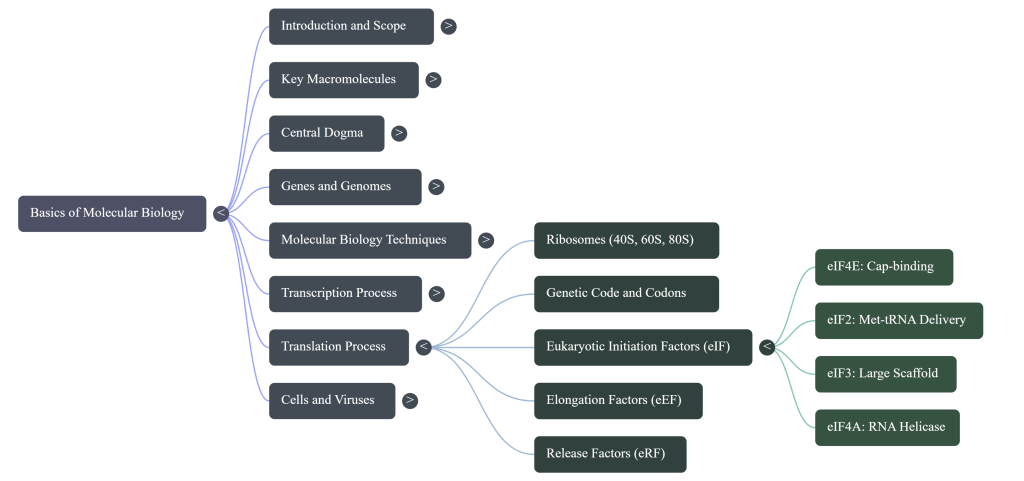
Translation takes place in the cytoplasm, where the cellular “factories” called ribosomes are located. These ribosomes consist of large and small subunits composed of proteins and ribosomal RNA (rRNA). Key participants include:
- mRNA: Acts as the template, carrying genetic information in nucleotide triplets called codons.
- tRNA (transfer RNA): Small molecules that transport specific amino acids to the ribosome. Each tRNA has an anticodon that is complementary to a specific mRNA codon, ensuring the correct amino acid is added to the growing chain,.
- Aminoacyl tRNA synthetase: The enzyme that catalyzes the bonding between a tRNA and its corresponding amino acid.
The Four Phases of Translation
Translation proceeds through four distinct phases to assemble a polypeptide chain:
- Activation: Before translation begins, a specific amino acid is covalently bonded to its correct tRNA, a process often referred to as “charging” the tRNA.
- Initiation: The small ribosomal subunit binds to the 5′ end of the mRNA with the help of initiation factors. It scans for the “start” codon (typically AUG, which codes for methionine) to begin the synthesis.
- Elongation: Amino acids are added one by one to the growing polypeptide chain. This involves positioning the correct tRNA in the ribosome, forming a peptide bond, and shifting the mRNA by one codon.
- Termination: The process ends when the ribosome encounters a stop codon (UAA, UAG, or UGA). Because no tRNA recognizes these codons, release factors intervene to cause the polypeptide chain to be released from the ribosome,.
Key Differences and Constraints
- Directionality: Translation occurs in the N -> C direction, meaning the first amino acid added forms the N-terminal of the protein.
- Energetics: The process is energy-intensive; translating a protein with n amino acids requires 4n−1 high-energy phosphate bonds.
- Speed: Translation is significantly faster in prokaryotes (up to 17–21 amino acids per second) than in eukaryotes (about 6–7 amino acids per second).
- Cellular Organization: In prokaryotes, translation and transcription can occur simultaneously in the cytoplasm,. In eukaryotes, transcription happens in the nucleus, and the mRNA must be processed and exported to the cytoplasm before translation can begin,.
Certain antibiotics (such as tetracycline or streptomycin) work by specifically targeting and inhibiting the translational machinery of bacterial cells without harming the host’s eukaryotic cells,.
7. Comparative Cellular Organization and Virology
Distinguishing between prokaryotic and eukaryotic architectures is vital for molecular research, as it dictates how genetic material is packaged and accessed.
Genomic Storage Comparison
- Prokaryotes: Generally possess a single large circular chromosome and plasmids (small, transferable DNA circles carrying traits like antibiotic resistance).
- Eukaryotes: Store DNA on multiple linear chromosomes packed with histones. These feature telomeres (repetitive caps) to prevent degradation and introns (non-coding regions) that are spliced out.
Virology and the Baltimore Classification
Viruses are classified by the Baltimore system into seven distinct classes based on their genetic contents and replication strategies:
- Class I: dsDNA viruses (e.g., T4 bacteriophage).
- Class II: ssDNA viruses (e.g., ϕX174).
- Class III: dsRNA viruses.
- Class IV: (+)-sense ssRNA viruses (e.g., MS2).
- Class V: (-)-sense ssRNA viruses.
- Class VI: ssRNA-RT viruses (retroviruses like HIV).
- Class VII: dsDNA-RT viruses.
The Viral Life Cycle:
- Attachment: Specific binding (e.g., HIV gp120 to CD4 receptors).
- Entry and Uncoating: Release of the viral genome into the host.
- Replication: Hijacking host machinery to synthesize viral components.
- Release: Exit via Lysis (rupture) or Budding (acquisition of an envelope).
8. Modern Molecular Techniques: Characterization and Manipulation
The ability to manipulate molecular components has revolutionized forensic science and the pharmaceutical industry.
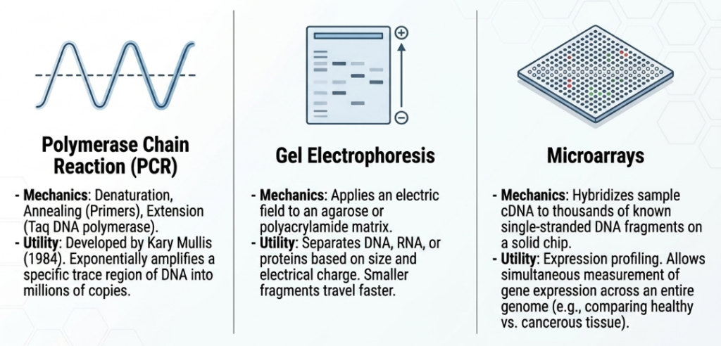
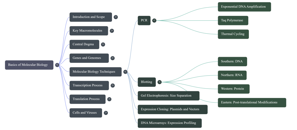
Polymerase Chain Reaction (PCR)
PCR utilizes a heat-stable Taq polymerase to exponentially amplify DNA.
- Mechanism: Thermal cycling separates strands (melting), allows primers to bind (annealing), and enables synthesis (extension).
- Applications: Genetic “fingerprinting,” diagnosis of infectious diseases, paternity testing, and phylogenetic analysis of ancient DNA.
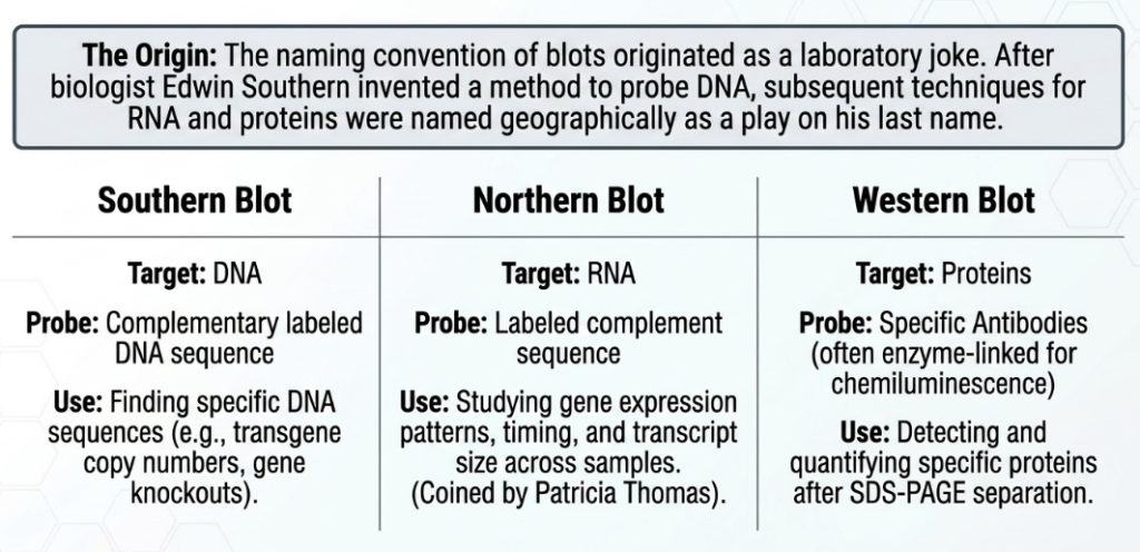
The Blotting Nomenclature
| Technique | Target Molecule | Primary Purpose |
|---|---|---|
| Southern | DNA | Probing for specific sequences or transgene copy numbers. |
| Northern | RNA | Analyzing gene expression patterns and levels. |
| Western | Protein | Quantitative analysis using specific antibodies. |
| Eastern | Protein | Detecting post-translational modifications. |
Advanced Manipulation
Expression Cloning uses plasmids to produce large quantities of proteins for drug testing or structural biology. DNA Microarrays enable expression profiling, allowing the simultaneous monitoring of thousands of genes to compare healthy and cancerous tissue.
8. Conclusion: The Future of Molecular Insights
Molecular biology remains anchored by the Central Dogma, yet it is continuously revised as we uncover the vast roles of non-coding RNA and the “continuum” of overlapping genes. The field has transitioned from viewing genes as discrete “beads on a string” to understanding them as part of a complex, overlapping regulatory landscape. The strategic value of molecular biology lies in its ability to decode life at its most fundamental level, providing the essential tools for both therapeutic innovation and our understanding of biological history.
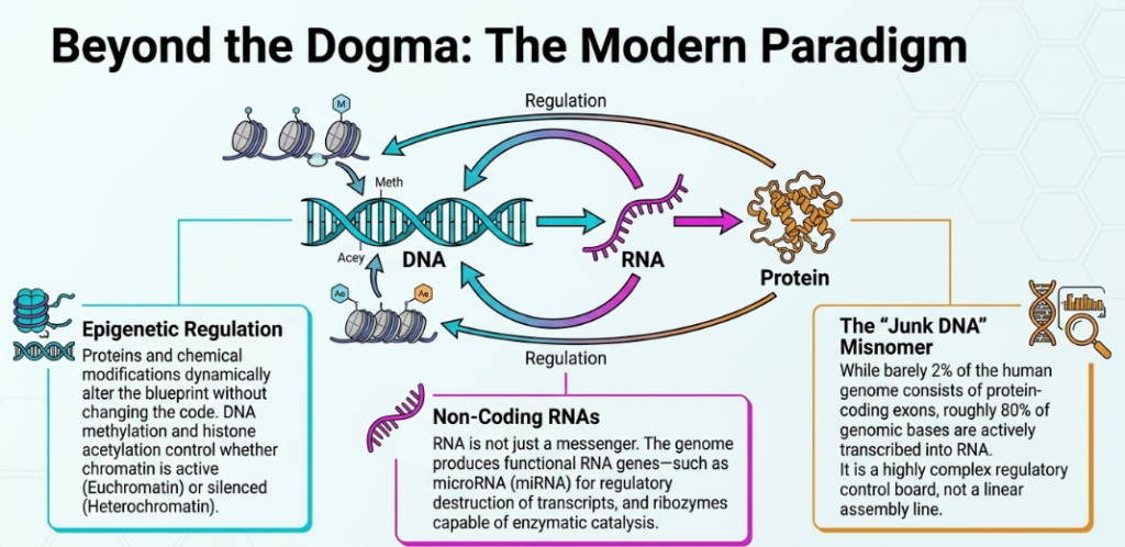
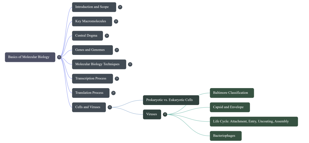
Image Summary
Questions/Answers
1. How do DNA, RNA, and proteins interact in the cell?
The interaction between DNA, RNA, and proteins is a fundamental concept in molecular biology, often described by the central dogma, which outlines a directional flow of genetic information: DNA is transcribed into RNA, which is then translated into proteins,. These three molecules interact through complex, highly regulated systems that ensure the cell functions, reproduces, and responds to its environment,.
The Flow of Information: Transcription and Translation
- DNA to RNA (Transcription): DNA serves as the primary repository of genetic information. When a gene is expressed, an enzyme called RNA polymerase (a protein) binds to a specific region of the DNA known as a promoter,. The RNA polymerase then creates an mRNA (messenger RNA) copy of the DNA sequence,. In eukaryotic cells, this occurs in the nucleus, and the resulting mRNA must be processed and exported to the cytoplasm before it can be used,.
- RNA to Protein (Translation): Once in the cytoplasm, the mRNA interacts with ribosomes, which are complex “factories” made of both proteins and ribosomal RNA (rRNA),. Transfer RNA (tRNA) molecules bring specific amino acids to the ribosome, matching their anticodons to the codons on the mRNA strand,. The ribosome then links these amino acids together to form a polypeptide chain, which folds into a functional protein,.
Proteins Regulating DNA and RNA
The relationship is not merely a one-way street; proteins are essential for managing and regulating the nucleic acids:
- Regulating Expression: Specific “regulative proteins” bind to DNA at the edges of genes to control their transcription into mRNA, effectively turning genes on or off,.
- DNA Packaging: In eukaryotic cells, DNA is wound around proteins called histones to form chromatin,. The way DNA is stored on these histones, and chemical modifications to the histones themselves, determine whether a region of DNA is accessible for transcription.
- Maintenance and Repair: A variety of proteins are responsible for DNA replication, unwinding the double helix and synthesizing new strands,. Other proteins monitor the genome for damage and perform repairs to maintain genetic integrity,.
Alternative Interactions and Functional RNA
While the DNA → RNA → protein path is standard, there are important variations:
- Reverse Transcription: Some viruses, such as retroviruses, can reverse this flow by using an enzyme to transcribe their RNA genome back into DNA,.
- Functional RNA: Not all RNA is intended for translation into protein. Some RNA molecules, such as ribozymes, have enzymatic functions, while others like miRNAs play direct regulatory roles in gene activity,.
- Splicing: After transcription, RNA can undergo splicing, where non-coding regions (introns) are removed and coding regions (exons) are joined. This process is catalyzed by the spliceosome, a large complex of proteins and small nuclear RNAs,.
2. Explain how PCR is used to amplify genetic material.
The Polymerase Chain Reaction (PCR) is a fundamental laboratory technique used to exponentially amplify specific fragments of DNA through in vitro enzymatic replication. Developed by Kary Mullis in 1984, it allows researchers to generate millions of copies of a target DNA sequence from even a single or trace amount of starting material.
The process relies on several key components and a repetitive cycle of temperature changes known as thermal cycling:
Essential Components
- DNA Template: The original sample of genetic material containing the specific region to be amplified.
- Heat-stable DNA Polymerase: Typically Taq polymerase, an enzyme isolated from the bacterium Thermus aquaticus that can withstand the high temperatures required to separate DNA strands.
- DNA Primers: Short synthetic oligonucleotides designed to be complementary to the ends of the target DNA region, providing a starting point for DNA synthesis.
- Nucleotides: The chemical building blocks (A, T, C, and G) used by the polymerase to construct the new DNA strands.
The Three-Step Thermal Cycle
PCR works by repeating a sequence of three temperature-controlled steps:
- Denaturation (Heating): The reaction mixture is heated to a high temperature to physically separate the double-stranded DNA into two single strands. This process is often called “DNA melting”.
- Annealing (Cooling): The temperature is lowered to allow the DNA primers to bind (anneal) to their complementary sequences on the single-stranded DNA templates.
- Extension (Synthesis): The DNA polymerase then assembles new DNA strands by adding nucleotides to the ends of the primers, using the single-stranded DNA as a template.
Exponential Amplification
As the cycle progresses, the DNA generated in one round serves as a template for the next round. This creates a chain reaction where the number of copies of the target region doubles with each cycle. After approximately 30 to 40 cycles, a single DNA fragment can be amplified into billions of copies.
Applications
Because of its high sensitivity and selectivity, PCR is indispensable for various applications, including:
- Medical Diagnostics: Detecting infectious diseases (like viruses or slow-growing bacteria) and diagnosing hereditary conditions.
- Forensics: Identifying genetic fingerprints from trace evidence or analyzing ancient DNA.
- Research: Isolating DNA fragments for sequencing, cloning, or functional gene analysis.
Reference
Li, Y., & Zhao, D. (2013). Basics of Molecular Biology.