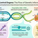
1. Introduction: The Instruction Manual for Life

Every living thing, from the smallest bacterium to the largest blue whale, is built and maintained using a detailed set of instructions. Think of this as a biological blueprint or a master instruction manual. This manual is a remarkable molecule called Deoxyribonucleic Acid (DNA). In a single human cell, this manual consists of about 3 billion letters of code. If you were to stretch the DNA from just one cell end-to-end, it would be about 2 meters long!

Found in nearly every cell of all living organisms, DNA contains the complete genetic information needed for development, function, growth, and reproduction. It is also the primary unit of heredity, responsible for passing traits from one generation to the next.

While this cellular manual is vast and complex, it is elegantly constructed from a few simple, repeating chemical parts. To understand how DNA works, we first need to look at its most fundamental building block.

2. The Basic Building Block: The Nucleotide
The entire DNA molecule is a polymer, which is simply a large structure made of many smaller, repeating units chained together. The fundamental repeating unit of DNA is called a nucleotide. Just as a wall is built from individual bricks, a DNA strand is built from a long chain of nucleotides.
Each DNA nucleotide, also known as a deoxyribonucleotide, consists of three core components:
1. A Five-Carbon Sugar (Deoxyribose): This sugar molecule forms a core part of the DNA’s structural framework.
2. A Phosphate Group: This group links the sugar of one nucleotide to the sugar of the next, forming a strong, continuous chain. These alternating sugar and phosphate units create the structural “sides” of the DNA ladder, known as the sugar-phosphate backbone.
3. A Nitrogenous Base: This is the most crucial component for information storage, as it contains the “letters” of the genetic code.

While every nucleotide has a sugar and a phosphate group, it is the nitrogenous base that gives each one its unique identity.

3. The “Rungs” of the Ladder: The Four Nitrogenous Bases
So if the backbone is always the same, where does the “code” come from? The answer lies in the nitrogenous bases. DNA uses four different bases to write its genetic code:
• Adenine (A)
• Guanine (G)
• Cytosine (C)
• Thymine (T)
These four bases belong to two distinct chemical families: the purines and the pyrimidines. The key difference between them is their size and structure.
| Base Family | Bases Included | Key Structural Feature |
| Purines | Adenine (A), Guanine (G) | Double-ring structure |
| Pyrimidines | Cytosine (C), Thymine (T) | Single-ring structure |
This difference in size is the secret to DNA’s elegant and consistent structure. To create a stable molecule, a two-ring purine always pairs with a single-ring pyrimidine. This consistent pairing ensures that the DNA “ladder” maintains a uniform width all along its length.

Now that we have identified all the individual parts, we can see how they are assembled to create the iconic final structure of DNA.
4. Assembling the Blueprint: The Double Helix
DNA is a double-stranded molecule, famous for its distinctive shape: the double helix. The most common and useful analogy for this shape is a twisted ladder. This structure is governed by three simple but powerful principles.
1. The Sugar-Phosphate Backbone The “sides” or “handrails” of the ladder are formed by the alternating sugar and phosphate components. The phosphate group of one nucleotide forms a strong covalent bond, known as a phosphodiester bond, with the sugar of the next nucleotide. This linkage repeats over and over, creating a continuous and durable chain.

2. Complementary Base Pairing The “rungs” of the ladder are formed by the nitrogenous bases. The two strands of DNA are held together in the middle by hydrogen bonds that form between the bases. As discovered by biochemist Erwin Chargaff, the amount of Adenine in any DNA molecule is always equal to the amount of Thymine, and the amount of Guanine always equals Cytosine. This crucial clue, known as Chargaff’s rules, led to the understanding of the following predictable pairing:
◦ Adenine (A) always pairs with Thymine (T), connected by two hydrogen bonds.
◦ Guanine (G) always pairs with Cytosine (C), connected by three hydrogen bonds. Because the G-C pair has three hydrogen bonds, it is stronger than the A-T pair. This variation in bond strength affects how DNA is replicated and transcribed, as regions with more A-T pairs are easier for cellular machinery to pull apart.

3. Antiparallel Strands The two DNA strands run in opposite directions relative to each other. This orientation is described as antiparallel. Imagine a two-lane highway where traffic flows in opposite directions. Similarly, one DNA strand runs in a 5′ (“five prime”) to 3′ (“three prime”) direction, while its complementary partner runs in the opposite 3′ to 5′ direction. These numbers refer to specific carbon atoms in the sugar ring. Crucially, they give each strand a direction, allowing the cellular machinery that ‘reads’ and ‘copies’ the DNA to know where to start and which way to go.

The genius of DNA’s design lies in how these principles work together. The strong sugar-phosphate backbone provides stability, while the weaker hydrogen bonds in the middle can be “unzipped” for copying. The strict base-pairing rules ensure that any copy is a perfect duplicate of the original, and the antiparallel strands provide the direction needed for this copying to happen efficiently. This creates a molecule that is both incredibly stable for storing information and easily replicable for passing it on.

It is this elegant architecture—two complementary, antiparallel strands twisted into a helix—that serves as the molecular foundation for all known life. This is the blueprint, copied and passed down through billions of years, that connects every organism on Earth.


Image Summary

Reference
Kumari, A. (n.d.). DNA: Deoxyribonucleic acid.
Travers, A., & Muskhelishvili, G. (2015). DNA structure and function. In FEBS Journal (Vol. 282, Issue 12, pp. 2279–2295). Blackwell Publishing Ltd. https://doi.org/10.1111/febs.13307
https://www.nature.com/scitable/topicpage/discovery-of-dna-structure-and-function-watson-397
https://openstax.org/books/concepts-biology/pages/9-1-the-structure-of-dna
https://www.nature.com/scitable/topicpage/introduction-what-is-dna-6579978
https://www.genome.gov/genetics-glossary/Deoxyribonucleic-Acid-DNA
https://en.wikipedia.org/wiki/DNA