1. Introduction to Neoplastic Capabilities
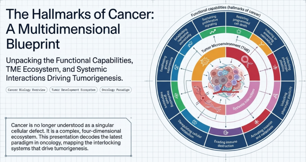
Cancer is fundamentally a complex, multi-stage evolutionary process characterized by the progressive acquisition of specific “Hallmarks of Cancer.” These functional capabilities serve as the definitive mechanistic operations required for tumorigenesis and malignant progression. The continuing premise of this framework is that these capabilities are not merely descriptive but are biological imperatives necessary—to varying degrees—in most forms of human cancer. To advance precision oncology, we must decode the “Dimensions of Cancer,” moving beyond the neoplastic cell to understand the broader ecosystem that sustains the disease state.
2. Dimension I: The Acquired Functional Capabilities (The Core Hallmarks)
The strategic importance of the nine discrete functional capabilities lies in their role as the primary outputs of a tumor’s evolutionary trajectory. It is critical to recognize that the acquisition of these hallmarks is strictly non-linear; dependencies on specific capabilities fluctuate across different cancer types and stages of development. A tumor may prioritize certain traits early in its life cycle while others become prominent only during late-stage dissemination.
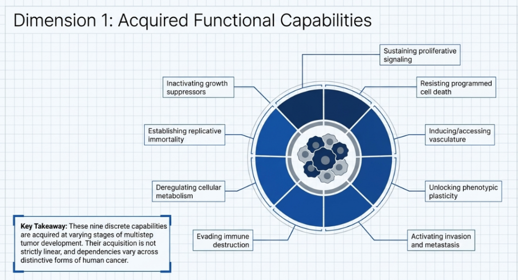
In the current synopsis, the functional capabilities are defined as follows:
1. Sustaining Proliferative Signaling: The chronic deregulation of growth-promoting instructions, often achieved through autocrine ligand production, subversion of stromal neighbors, or constitutive activation of downstream transducers like B-Raf or PI3-kinase.
- Neoplastic growth is fueled by diverse mechanisms that achieve growth factor independence:
- Autocrine Stimulation: Cancer cells produce growth factor ligands to which they respond via cognate receptors, creating self-sustaining loops.
- Receptor Hyper-responsiveness: Upregulation of receptor proteins at the cell surface or structural alterations allows for ligand-independent firing.
- Constitutive Transducer Activation: Somatic mutations in downstream transducers, such as B-Raf (~40% of melanomas) or PI3-kinase circuitry, ensure that proliferative signals remain “on” regardless of external ligand availability.
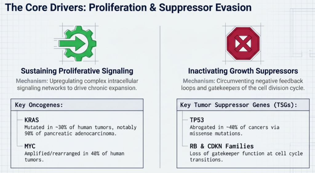
2. Evading Growth Suppressors: The circumvention of powerful programs—prototypically governed by RB (gatekeeper of the cell cycle) and TP53 (guardian of the genome)—that maintain tissue homeostasis.
- To maintain expansion, tumors must bypass “stop” signals governed by tumor suppressors. This deconstruction focuses on the central “gatekeeper” nodes, RB and TP53.
| Regulatory Node | Biological Input | Strategic Function | Pathway Impact |
|---|---|---|---|
| RB (Retinoblastoma) | Diverse extracellular and intracellular signals. | Integrates signals to decide on cell-cycle entry. | Loss permits persistent, inappropriate progression through the G1/S checkpoint. |
| TP53 (Guardian of the Genome) | Intracellular stress (DNA damage, hypoxia, low glucose). | Triggers growth arrest or apoptosis in response to suboptimal conditions. | Inactivation disables the primary sensor for irreparable genomic damage, enabling survival of unfit genotypes. |
3. Resisting Cell Death: The attenuation of apoptosis and the strategic use of autophagy to support survival under metabolic or therapeutic stress.
The “apoptotic trigger” is a high-stakes rheostat controlled by the Bcl-2 family. The balance between pro-apoptotic proteins (Bax and Bak) and anti-apoptotic relatives (Bcl-2, Bcl-xL) determines mitochondrial membrane integrity. Cancer cells strategically shift this balance to prevent the release of cytochrome c, thereby silencing both extrinsic and intrinsic death programs.
The Dual Role of Autophagy
Autophagy functions as a complex survival module:
- Tumor Barrier: In early stages, it acts as a suppressive mechanism by maintaining cellular homeostasis.
- Cytoprotective Survival: In established tumors, it serves as a nutrient-recycling program that allows stressed cells to enter a state of reversible dormancy, facilitating survival through the “metabolic desert” of chemotherapy.
4. Enabling Replicative Immortality: The maintenance of telomeric DNA via telomerase (TERT) or alternative recombination-based mechanisms to bypass senescence and crisis.
While normal cells enter senescence or crisis due to telomere erosion, cancer cells upregulate telomerase (TERT) to maintain chromosomal ends. Strategic analysis reveals that TERT provides more than just DNA maintenance; its noncanonical functions include:
- Wnt Pathway Amplification: TERT acts as a cofactor for the β-catenin/LEF transcription factor complex, directly fueling proliferative signaling.
- Enhanced Survival: Direct resistance to apoptosis and participation in DNA-damage repair independent of telomere length.
5. Inducing or Accessing Vasculature: The activation of the “angiogenic switch” or the co-option of existing normal tissue vessels to satisfy the metabolic demands of expanding neoplastic growths.
The “angiogenic switch” is typically tripped early in the premalignant phase. It is a biological rheostat governed by the balance of inducers (e.g., VEGF−A) and inhibitors (e.g., TSP−1). Critically, the tumor recruits a repertoire of bone marrow-derived cells—including macrophages, neutrophils, mast cells, and myeloid progenitors—to the TME margins. these cells provide the paracrine support necessary to sustain a leaky, convoluted, and erratic neovasculature.
6. Activating Invasion and Metastasis: The initiation of the invasion-metastasis cascade, frequently co-opting developmental programs like the epithelial-to-mesenchymal transition (EMT) to facilitate systemic dissemination.
The “Invasion-Metastasis Cascade” involves local invasion, intravasation, transit, and colonization. This process is choreographed by the Epithelial-to-Mesenchymal Transition (EMT), where cells lose their polygonal epithelial constraints for a spindly, fibroblastic morphology. This program is driven by pleiotropic transcription factors: Snail, Slug, Twist, and Zeb1/2.
Strategic Plasticity: EMT and MET
Invasion is not a one-way street. Disseminated cells often lose the stromal signals that induced EMT. This allows for a Mesenchymal-to-Epithelial Transition (MET), enabling the cell to revert to a noninvasive state to colonize distant tissues, effectively recapitulating the histopathology of the primary tumor in a new environment.
7. Reprogramming Cellular Metabolism: The major deregulation of cellular energetics and biosynthetic pathways to support continuous growth and proliferation.
8. Evading immune destruction: Orchestrating an immunosuppressive environment to circumvent detection and elimination by T cells and other immune effectors.
9. Unlocking phenotypic plasticity: Escaping committed states of differentiation, allowing cancer cells to adopt multiple phenotypic substates for survival and adaptation.
- Three Modes of Disrupted Differentiation
- Dedifferentiation: Mature cells “reverse course” to return to a progenitor-like state.
- Example: In colon cancer, the loss of transcription factors HOXA5 and SMAD4 allows mature epithelial cells to regress into invasive, stem-like states.
- Blocked Differentiation: Progenitor cells are “frozen” in an immature, highly proliferative stage.
- Example: In Acute Promyelocytic Leukemia (APL), the PML-RARα fusion protein traps myeloid cells in a proliferative state, preventing them from becoming mature granulocytes.
- Transdifferentiation (Lineage Switching): Cells switch from their preordained program to an entirely different one.
- Dedifferentiation: Mature cells “reverse course” to return to a progenitor-like state.

The “So What?” Layer
These capabilities transform a quiescent cell into a “chronically expansive” disease state. Because acquisition is non-linear, a tumor’s reliance on a specific hallmark may be transient, complicating therapeutic efforts. However, since these traits are functionally necessary for most human cancers, they provide a rational basis for categorizing the diverse pathology of neoplasia into a unified mechanistic framework.
3. Dimension II: Enabling Phenotypic Characteristics
Enabling characteristics are not functional end-goals themselves; rather, they are instrumental traits that facilitate the acquisition of hallmark capabilities. They represent the genomic and physiological “instability” that drives tumor evolution.

The five recognized enabling characteristics include:
1. Loss of genomic integrity: The foundational enabler involving gene mutations, Chromosomal Instability (CIN), and the presence of extrachromosomal DNA (ecDNA). The breakdown of “caretaker” machinery (DNA repair, damage sensing) increases the mutation rate. This provides the genetic substrate for the succession of clonal expansions, where the fittest mutant genotypes dominate.

2. Non-mutational epigenetic reprogramming: A separable enabling trait “markedly distinct from mutational cancer genetics” involving the dynamic regulation of gene expression. Independent of DNA mutation, the genome can be reprogrammed by microenvironmental stresses. Hypoxia can reduce the activity of TET demethylases, leading to global hypermethylation. Additionally, ECM stiffness triggers neuronal signaling circuits (glutamate/NMDAR) that induce invasiveness. This epigenetic heterogeneity creates a “mixed population” that enhances overall tumor fitness and therapeutic resistance. A notable example is the ZEB1/SETD1B feedback loop, where the TF ZEB1 induces a histone methyltransferase (SETD1B) that sustains the invasive EMT state through a self-reinforcing epigenetic circuit.
Tumor Microenvironment (TME) Triggers of Epigenetic Change
The physical and chemical environment of the tumor forces these software changes through three primary triggers:
- Hypoxia (Low Oxygen): Insufficient blood flow reduces the activity of TET demethylases, causing hypermethylation of the genome and silencing protective genes.
- Mechanical Stiffness: As the extracellular matrix (ECM) becomes dense, the resulting “stiffness” triggers gene-expression networks via integrin receptors, eliciting invasive hallmark traits.
- Paracrine Signaling: Nearby stromal cells release soluble factors that induce the Epithelial-to-Mesenchymal Transition (EMT), granting cancer cells migratory powers.
3. Polymorphic microbiomes: The influence of local and systemic microbial populations on tumor progression and treatment response. The microbiome is now established as a discrete enabling characteristic that modulates hallmarks across various barrier tissues (skin, lung, gut).
- Mutagenesis: Specific strains, such as PKS+ E. coli, produce toxins that directly damage host DNA, contributing to genome instability.
- Immune Modulation: Microbiota can circulate systemic factors that modulate adaptive immunity. For instance, Enterococcus strains expressing the SagA peptidoglycan hydrolase release mucopeptides that activate the NOD2 receptor, enhancing T-cell responses. Conversely, bacterial-produced inosine serves as a rate-limiting metabolite that can further modulate T-cell activity.
- Metabolic Influence: Butyrate-producing bacteria have pleiotropic effects, including the induction of senescence in some contexts and the modulation of energy metabolism to promote tumorigenesis in others.
Mechanisms of Microbial Influence
| Mechanism of Influence | Biological Effect | Resulting Hallmark Capability |
|---|---|---|
| Mutagenesis | Production of toxins (e.g., E. coli PKS locus). | Genome Instability & Mutation |
| Metabolic Signaling | Secretion of metabolites like butyrate. | Tumor-Promoting Inflammation |
| Immune Modulation | Mucopeptides (e.g., SagA) activating NOD2. | Avoiding Immune Destruction |
The Intratumoral Microbiota
One of the most provocative discoveries is that bacteria don’t just live on the surface of tissues; they exist inside the tumor. Specific bacteria live within cancer cells and immune cells, directly influencing the tumor’s phenotype, growth, and how it responds to chemotherapy.
The “So What?”: The microbiome is now recognized as a quasi-independent variable in oncology. A patient’s response to immunotherapy may depend more on their microbial “tenants” than their own genetics.
Transition: The inflammatory stress caused by dysbiosis often results in the accumulation of “zombie-like” senescent cells.
4. Tumor-promoting inflammation: Paradoxically, immune cells recruited to scan for damage supply the TME with growth factors and matrix-degrading enzymes. This “inflammation” inadvertently fuels hallmark functions, such as angiogenesis and invasion.
5. Innervation: The active recruitment of various neuronal subtypes and their axonal projections into the neoplastic microenvironment.
The “So What?” Layer
The “Loss of genomic integrity” acts as the engine of cancer’s expansive nature. This instability—fueled by telomere erosion and defects in DNA damage-sensing and repair pathways (such as mismatch repair and homologous recombination)—often results in the generation of linear and circular ecDNA. These ecDNA elements are critical drivers of oncogene overexpression (e.g., MYC, EGFR), providing the genetic diversity required for tumors to evade therapy and adapt to environmental stress.
4. Dimension III: Hallmark-Conveying Cells of the Tumor Microenvironment (TME)
In modern systems biology, a tumor is viewed as an “outlaw organ” or a “cellular ecosystem.” This ecosystem is populated by heterotypic cells that are recruited, reprogrammed, and corrupted to act as “accessories to the crime,” facilitating hallmark manifestation.
| Cell Type | Role in Facilitating Hallmarks | Example Mechanism |
|---|---|---|
| Cancer-associated fibroblasts (CAFs) | Structural and metabolic support | Secreting HGF/TGF-β to promote growth and invasion |
| Macrophages and neutrophils | Immunosuppression and remodeling | Secretion of IL-10 and VEGF to aid evasion and angiogenesis |
| Endothelial cells and pericytes | Vascular homeostasis | Formation of leaky, irregular vessels via VEGFR signaling |
| Neurons and axons | Neurotrophic support | Releasing neurotransmitters that trigger proliferative signaling |
| Senescent cells | Pro-tumorigenic secretome | Promoting a SASP that alters the TME to favor neighboring cancer cells |

Senescent Cells: Functional Integration into the Tumor Microenvironment
Historically viewed as a protective barrier to prevent neoplasia, cellular senescence is now recognized as a tumor-promoting liability in the malignant microenvironment.
The Senescence-Associated Secretory Phenotype (SASP) is the primary mechanism through which senescent cells provide paracrine support for malignancy. By releasing a complex repertoire of cytokines, chemokines, and proteases, senescent cells contribute to proliferative signaling, angiogenesis, and immunosuppression.
Critically, recent data necessitate a focus on transitory or reversible senescence. Senescent cancer cells may “exit” this state to resume proliferative expansion—a phenomenon known as senescence exit. This serves as a vital mechanism for therapy resistance and dormancy, allowing cancer cells to survive cytotoxic treatment before re-emerging as more aggressive, viable clones. The senescent population in the tumor microenvironment (TME) is heterogeneous, originating from cancer cells, Cancer-Associated Fibroblasts (CAFs), and endothelial cells, all of which are functionally integrated into the “outlaw organ” ecosystem.
The SASP: The “Zombie” Factory
Senescent cells develop a Senescence-Associated Secretory Phenotype (SASP), pumping out cytokines and proteases that convey hallmark capabilities to nearby viable cancer cells in four ways:
- Inducing Angiogenesis: Stimulating blood vessel growth.
- Sustaining Proliferative Signaling: Sending growth signals to neighbors.
- Stimulating Invasion: Using proteases to break down tissue barriers.
- Evasion of Immunity: Creating an immunosuppressive “shield.”
The “So What?” Layer
The strategic significance of the TME lies in the realization that virtually all accessory cells exist in multiple phenotypic substates. These cells are “reprogrammed” by the tumor to contribute to its survival. Consequently, the tumor functions as an integrated, multi-cellular unit rather than a simple mass of malignant clones, necessitating therapies that disrupt these heterotypic dependencies.
5. Dimension IV: Multifaceted Systemic Interactions
Beyond the local microenvironment, cancer is governed by Dimension IV: systemic parameters that influence hallmark acquisition and disease progression. Factors such as Aging and Obesity are now recognized as critical systemic modifiers. Aging contributes to the accumulation of senescent cells and the erosion of immune surveillance, while obesity drives systemic inflammation and metabolic shifts that facilitate the “Deregulating cellular metabolism” hallmark. These systemic interactions define the physiological context in which the other three dimensions operate.
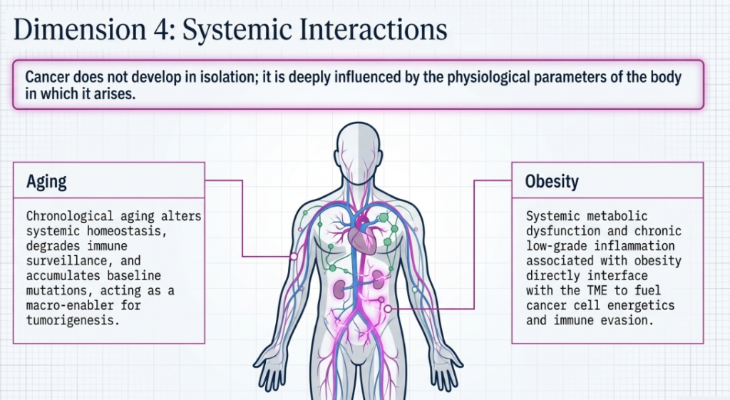
6. Molecular Orchestrators: The Roles of Oncogenes and Tumor Suppressors
At the molecular level, hallmark capabilities are often anchored by genetic “drivers.” Current research, specifically reflected in the COSMIC database, has identified 193 defined proliferation-driving oncogenes that sustain the chronically expansive state of the disease.
- KRAS: Mutated in ~30% of human tumors. It is nearly ubiquitous in pancreatic adenocarcinoma (90%) and highly prevalent in colorectal cancer (50%) and lung adenocarcinoma (35%).
- MYC: A master transcription factor (TF) regulating thousands of genes. It is amplified or rearranged in 40% of human tumors, often driven by ecDNA-mediated overexpression.
- TP53: The primary “gatekeeper” of the cell cycle, mutated in ~40% of cancers. Crucially, 60%–70% of these are missense mutations centered on hotspots, abrogating its ability to sense DNA damage and trigger repair or apoptosis.
The “So What?” Layer
While these molecular orchestrators are potent, a single mutation (e.g., in KRAS) is evidently not sufficient for full tumorigenesis. Malignant progression requires a multi-step synergy of genetic alterations, epigenetic reprogramming, and the corruption of the TME, reinforcing the need for a multidimensional diagnostic approach.
7. Clinical Synthesis: Therapeutic Targeting of the Hallmarks
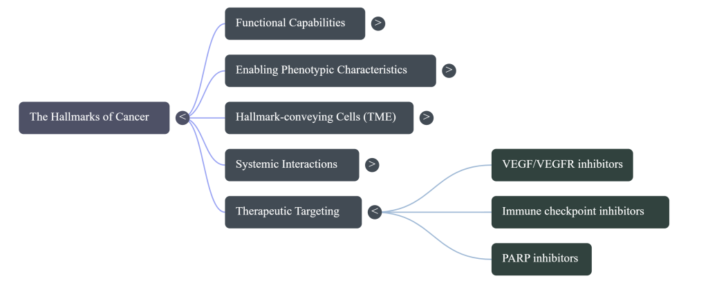
The clinical shift from cytotoxic “carpet-bombing” to mechanism-targeted therapy represents the practical application of the hallmark framework. By co-targeting multiple functional capabilities, we can overcome the resilience of the cancer ecosystem.
Current and emerging therapeutic strategies include:
- VEGF/VEGFR inhibitors: Disrupting the “Inducing or accessing vasculature” hallmark.
- Immune checkpoint inhibitors: Reversing the “Evading immune destruction” capability by restoring T-cell activity.
- PARP inhibitors: Exploiting the “Loss of genomic integrity,” specifically targeting tumors with DNA damage-repair defects such as BRCA mutations.

The “So What?” Layer
The future of oncology lies in addressing the complexity of the “outlaw organ.” This involves shifting from killing cancer cells in isolation to “reprogramming” immunosuppressive cell types within the TME, effectively turning the tumor’s supportive ecosystem against itself.
8. Conclusion and Key Takeaways
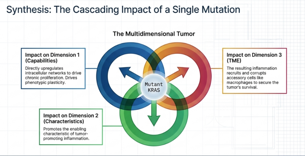
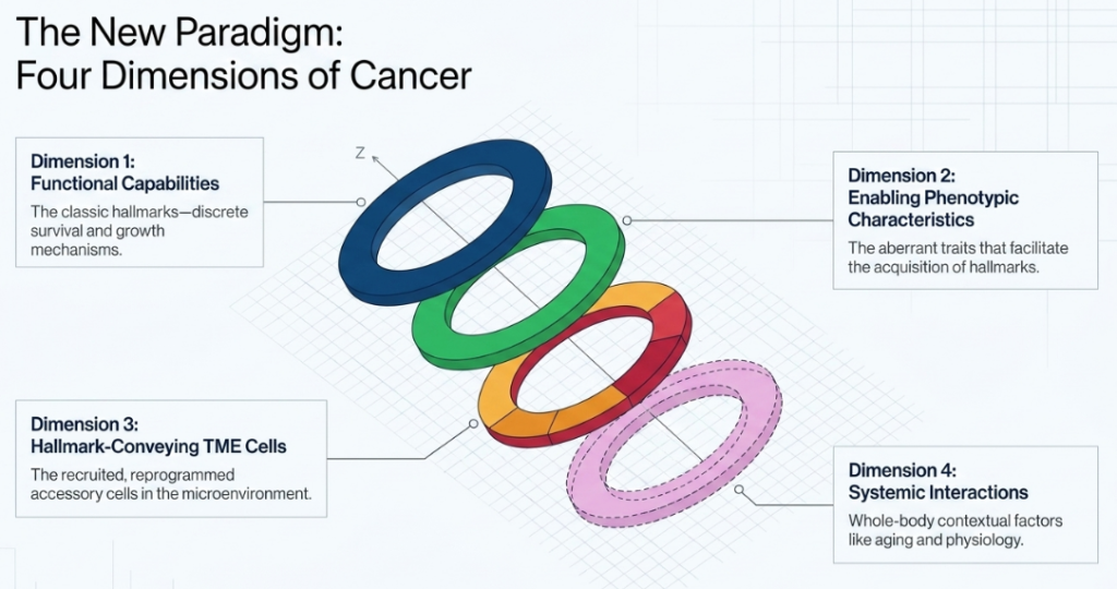
Cancer is defined by four distinctive conceptual dimensions: Functional capabilities, Enabling traits, Hallmark-conveying TME cells, and Systemic interactions. This framework establishes that a tumor is a sophisticated “cellular ecosystem” that corrupts physiological processes to sustain its own growth. Understanding the interplay between these dimensions—from ecDNA-driven genomic disarray to the systemic influences of obesity and aging—is essential for the next generation of mechanism-targeted oncology.
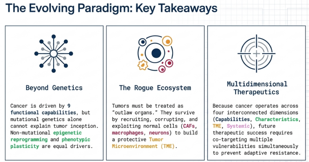
Image Summary:


References
Hanahan, D., & Weinberg, R. A. (2000). The Hallmarks of Cancer. In Cell (Vol. 100).
Hanahan, D., & Weinberg, R. A. (2011). Hallmarks of cancer: The next generation. In Cell (Vol. 144, Number 5, pp. 646–674). https://doi.org/10.1016/j.cell.2011.02.013
Hanahan, D. (2022). Hallmarks of Cancer: New Dimensions. In Cancer Discovery (Vol. 12, Number 1, pp. 31–46). American Association for Cancer Research Inc. https://doi.org/10.1158/2159-8290.CD-21-1059
Hanahan, D. (2026). Hallmarks of cancer—Then and now, and beyond. In Cell (Vol. 189, Number 8, pp. 2254–2277). Elsevier B.V. https://doi.org/10.1016/j.cell.2025.12.049


Related posts:
 Epstein-Barr Virus Protein EBNA1 Found to Boost Cancer-Linked Genes in HeLa Cells
 Natural Healers: Animals That Cure Themselves with Natural Solutions
 How Neuronal Activity Fuels Small Cell Lung Cancer: A New Pathogenesis Pathway
 How Fluctuating DNA Methylation Maps Cancer Evolution in Real Time
 Smart Biosensors: Dual ACE2 Epitope Receptors Offer Rapid Detection of SARS-CoV Variants