A Hidden Cellular Mechanism Comes to Light
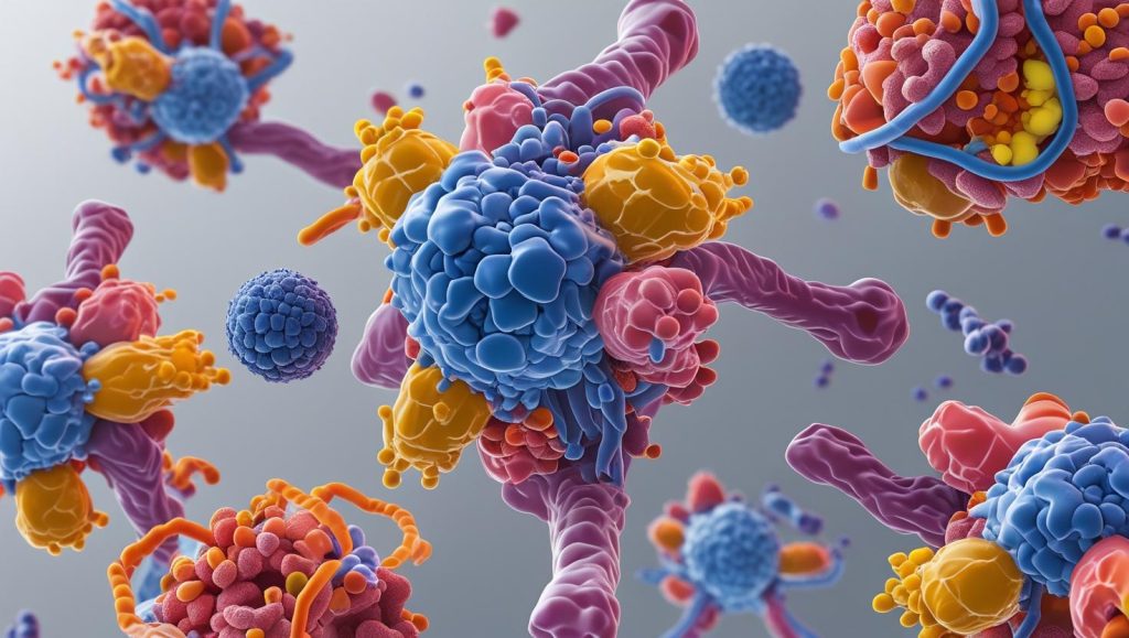
In a breakthrough discovery reshaping our understanding of how cells manage internal cargo, researchers at the NIH and University of Virginia have identified a new class of vesicle complexes called hemifusomes. These structures, captured in living cells via advanced cryo-electron tomography (cryo-ET), challenge the long-standing model of multivesicular body (MVB) formation dominated by ESCRT protein complexes.
Instead of relying on canonical budding mechanisms, hemifusomes use long-lived hemifusion diaphragms and proteolipid nanodroplets (PNDs) to build intraluminal vesicles — a process the authors propose as an ESCRT-independent route for MVB biogenesis.
What Are Hemifusomes?
Hemifusomes are organelles formed by two vesicles in a unique configuration:
- Direct hemifusomes: A smaller vesicle hemifused to the cytoplasmic side of a larger one.
- Flipped hemifusomes: The smaller vesicle is embedded within the lumen of the larger one.
Both forms share a hemifusion diaphragm (HD)—a thin membrane layer that connects two vesicles without fully merging their contents. These HDs were previously thought to be transient and unstable. This study, however, reveals HDs up to 160 nm in size that remain intact for extended periods, acting as a platform for vesicle development and cargo sorting.
“These aren’t just fleeting intermediates. Hemifusomes are real, stable, and active participants in membrane remodeling,” says co-author Dr. Seham Ebrahim.
The Power of Proteolipid Nanodroplets
The formation of hemifusomes appears to be initiated or stabilized by proteolipid nanodroplets (PNDs)—dense, protein-lipid structures about 42 nm in size that nestle into the membrane rim where the HD forms.
These nanodroplets likely supply the membrane material and protein cargo required to generate new vesicle compartments, acting as localized hubs of vesiculogenesis—a term the authors use to describe de novo vesicle formation from within the membrane.
Why This Matters: Challenging the ESCRT Paradigm
The conventional model of intraluminal vesicle formation within MVBs involves the ESCRT (Endosomal Sorting Complex Required for Transport) machinery. But the hemifusome mechanism operates without this system:
- No omega-shaped budding necks typical of ESCRT vesicles.
- No uptake of gold nanoparticle tracers used to label endocytic cargo.
- Distinct luminal contents compared to canonical endosomes or lysosomes.
This suggests hemifusomes represent an entirely new vesicle biogenesis pathway, with significant implications for understanding cargo sorting, membrane recycling, and even disease processes linked to defective vesicle trafficking.
Visualizing the Invisible
Thanks to cryo-ET, the researchers were able to capture high-resolution, 3D images of hemifusomes in four different mammalian cell types. Their unique topology—flat lens-shaped internal vesicles and variable fusion angles—was previously invisible using conventional electron microscopy due to preparation artifacts.
These observations also offer physical validation of membrane models predicted in computational simulations but never seen in living cells—until now.
Rewriting the Vesicle Lifecycle
Perhaps most strikingly, hemifusomes appear to:
- Participate in cargo sorting and membrane recycling,
- Act as precursors to multivesicular bodies, and
- Operate as self-organizing vesicle factories in certain cellular zones.
This new mechanism may complement or even compete with ESCRT pathways, especially in conditions where ESCRT machinery is deficient or compromised.
What’s Next?
Researchers plan to:
- Identify the molecular triggers that initiate hemifusome formation.
- Determine if these structures play roles in neurological disorders or cancer.
- Explore if proteolipid nanodroplets can be harnessed for targeted drug delivery or synthetic biology applications.
TL;DR:
Scientists have uncovered “hemifusomes”—a new type of vesicle complex stabilized by proteolipid nanodroplets. This discovery introduces a completely new, ESCRT-independent route for multivesicular body formation and reveals deeper layers of complexity in how cells handle internal cargo.
Reference:
Tavakoli, A., Hu, S., Ebrahim, S., & Kachar, B. (2025). Hemifusomes and interacting proteolipid nanodroplets mediate multi-vesicular body formation. Nature Communications, 16(1), 1-15. https://doi.org/10.1038/s41467-025-59887-9
Related posts:
 New Class of Antibiotics with a Novel Mode of Inhibition against World Health Organization’s Critical Listed Bacteria, Carbapenem-Resistant Acinetobacter baumannii
 Development of a Highly Efficient Cancer Treatment Strategy using the Vibrionic-Driven Action of Synthetic Aminocyanines
 Natural Healers: Animals That Cure Themselves with Natural Solutions
 Whats in your Gut? One ingestible pill coming up for understanding the microbiota inside small intestine
 Bacterial Cellulose: A Game-Changer for Plant Wound Healing and Regeneration

