Introduction: The Revolutionary Gene-Editing Tool
Imagine you could edit the source code of a living organism just like you edit a document on a computer. CRISPR-Cas9 is a revolutionary technology that makes this possible, acting like a biological “Find and Replace” function for DNA. This powerful and precise tool allows scientists to make specific changes to the genome—the complete set of genetic instructions—of an organism.

To begin, let’s explore where this incredible technology originated.
The Origin Story: Nature’s Smartest Immune System
The story of CRISPR doesn’t begin in a high-tech laboratory but in the sun-baked salt marshes of Santa Pola, Spain. In 1989, a graduate student named Francisco Mojica was studying the salt-loving microbe Haloferax mediterranei when he stumbled upon a strange, repeating pattern in its DNA. Captivated by this molecular puzzle, he dedicated years to understanding what he had discovered: a natural marvel, an ancient and elegant immune system that bacteria use to fight off invading viruses. This system, now known as CRISPR, acts as a genetic memory, allowing bacteria to recognize and destroy their viral enemies. This article will explain how scientists ingeniously repurposed this natural bacterial defense into CRISPR-Cas9, one of the most powerful and accessible gene-editing tools in history.

Think of it like a molecular vaccination card. When a virus attacks, the bacterium “remembers” the invader by storing a piece of its DNA. If that same virus tries to attack again, the bacterium can quickly recognize and destroy it. Its defense strategy unfolds in three elegant stages, creating a specific and heritable immune memory.
1. Adaptation: The “Mugshot” The bacterium’s first move is to capture a small piece of the invader’s DNA. This viral DNA fragment is then integrated into a special region of the bacterium’s own genome called the CRISPR locus. Here, it is stored as a unique “spacer” sequence, nestled between repeating DNA segments. These spacers, derived directly from viral genomic sequences, act as a molecular “mugshot” of the enemy, forming an immune memory that is passed down to all the bacterium’s offspring.
2. Expression: The “Wanted Poster” Next, the CRISPR locus, now containing a library of viral mugshots, is transcribed into a long RNA molecule. This molecule is then processed into smaller pieces called CRISPR RNAs (crRNAs). Each crRNA contains the sequence of a single spacer—a molecular “wanted poster” for a specific virus. In the Type II CRISPR system (the one famously adapted for gene editing), each crRNA teams up with a second essential RNA called tracrRNA to form a functional surveillance unit.
3. Interference: The “Security Guard” Finally, the crRNA guide, along with its tracrRNA partner, binds to a Cas9 protein. This complex is the “security guard” of the cell. It patrols the bacterium’s interior, searching for any foreign DNA. When it encounters DNA that perfectly matches the sequence on its crRNA “wanted poster,” the Cas9 protein locks on and makes a precise, double-stranded cut. This single snip is all it takes to neutralize the viral DNA, stopping the infection in its tracks.

“CRISPR provides acquired resistance against viruses in prokaryotes.”
Scientists brilliantly adapted this ancient bacterial defense mechanism, transforming it from a tool for fighting viruses into a programmable tool for editing the DNA of nearly any organism, including plants.
How It Works: The Gene Editor’s Toolkit
Researchers brilliantly simplified the natural bacterial system into a versatile, two-part toolkit that can be used to edit genes in plants, animals, and even humans.
• The “Molecular Scissors” (Cas9 protein): Cas9 is a protein known as a nuclease. Its job is simple but powerful: to cut DNA. Guided by its RNA partner, Cas9 makes a precise, clean, double-strand break in the DNA’s helix structure.
• The “GPS Navigator” (Guide RNA): In nature, two separate RNA molecules (crRNA and tracrRNA) are needed to guide Cas9. Scientists streamlined this by fusing them into a single, easy-to-make molecule called a guide RNA (gRNA). Simplifying the natural three-part system into this two-part version was a crucial innovation that made the technology vastly more practical, scalable, and accessible for thousands of labs worldwide. About 20 letters of this gRNA are a customizable sequence that acts like a GPS coordinate, telling the Cas9 scissors exactly where to go and make a cut in the vast genome.
For the system to work, one more crucial element is needed: the PAM sequence. The PAM (Protospacer Adjacent Motif) is a short, specific DNA sequence (for the commonly used Cas9, this is NGG) that must be located right next to the target DNA. You can think of it as a “molecular handle” that Cas9 must grab onto before it can make its cut. This PAM requirement is a clever safety mechanism inherited from the bacterial immune system, preventing Cas9 from cutting the bacterium’s own CRISPR locus, which contains the viral memories but lacks the PAM sequence.
With the scissors loaded with their GPS coordinates, the system was ready to be sent into a cell to find its target and make the first, critical cut.
The Key Players
The system can be thought of as a biological “search and cut” tool, with three essential parts that work in concert.

| Component | Function & Analogy |
|---|---|
| Cas9 Nuclease | The “molecular scissors.” A protein enzyme that precisely cuts both strands of the target DNA. Its nuclease domains (HNH and RuvC) execute the cut. |
| Single-guide RNA (sgRNA) | The “GPS” or programmable guide. Scientists engineered this single RNA by fusing two of nature’s components (crRNA and tracrRNA). Its sequence is designed to match a target gene, guiding the Cas9 scissors to the exact right spot in the genome. |
| Protospacer Adjacent Motif (PAM) | The “docking site” or molecular checkpoint. A short, specific DNA sequence (for the common S. pyogenes Cas9, this is 5′-NGG-3′) that Cas9 must recognize on the target DNA before it can make its cut. |
The Editing Process: A Step-by-Step Guide
For gene editing to occur, the Cas9 protein and the custom-designed sgRNA must first be delivered into the nucleus of a plant cell, where they combine to form a complex. From there, the process unfolds in three main steps.
Step 1: Finding the Target
The sgRNA component acts as the navigator. The complex scans the plant’s vast genome, searching for the one location where the DNA sequence is perfectly complementary to the guide RNA’s sequence.
Step 2: The Secret Handshake (PAM Sequence)
Before it can make a cut, the Cas9 protein needs to verify the location with a “secret handshake.” It looks for a specific, short DNA sequence called the Protospacer Adjacent Motif (PAM). This PAM sequence must be located immediately next to the target DNA. For the commonly used Cas9 system, this sequence is 5′-NGG-3′. The PAM acts as a critical checkpoint; if Cas9 doesn’t find it, it will not bind or cut the DNA, even if the sgRNA has found its match.
Step 3: Making the Cut (Double-Strand Break)
Once the sgRNA has bound to the target DNA and Cas9 has recognized the PAM sequence, the molecular scissors are activated. Cas9 uses two separate nuclease domains, known as HNH and RuvC, to make a clean cut across both strands of the DNA double helix. This creates a double-strand break (DSB). The creation of this DSB is the central event in CRISPR editing—it’s a deliberate break in the DNA that signals the cell to begin repairs.
The edit itself doesn’t happen when Cas9 makes the cut. The actual genetic modification occurs in the next stage, when the plant cell uses its own natural repair machinery to fix the damage.
After the Cut: How the Cell Repairs and Edits its DNA
Once the DNA is broken, the plant cell immediately activates its own repair systems to mend the break. The outcome of the gene edit depends entirely on which of the two primary repair pathways the cell uses.
◦ Non-Homologous End Joining (NHEJ): This is the cell’s default, fast-acting, but somewhat messy repair crew. It stitches the two broken ends of the DNA back together. In the process, it often makes small errors, creating tiny insertions or deletions of DNA letters (called “indels”). While an “error” for the cell, this is incredibly useful for scientists, as these indels can effectively disrupt or “knock out” a specific gene.
◦ Homologous Recombination (HR): This is the cell’s more precise, but much less common, repair pathway. If scientists provide a DNA template alongside the CRISPR system, the cell can use this template to repair the break. This allows for a “precise gene insertion,” correction, or replacement.

The table below compares these two crucial pathways.
| Feature | Non-Homologous End Joining (NHEJ) | Homology-Directed Repair (HR) |
|---|---|---|
| Analogy | “Quick & Dirty” Emergency Crew | “Precision Repair Crew” with a Blueprint |
| Mechanism | The cell’s machinery directly glues the two broken ends of the DNA back together without a guide. It is a fast but often imperfect “emergency” repair. | The cell’s machinery uses a separate piece of DNA, called a donor template, as a blueprint to precisely repair the break. It is a slower but highly accurate “precision” repair. |
| Outcome | This pathway often introduces small errors, such as adding or deleting a few DNA letters (called “indels”). These errors can disrupt a gene’s function, effectively turning it off. This is known as a gene knockout. This is useful for studying what a gene does or for disabling a gene that causes a plant disease. | Because it follows a template, this pathway can be used to make precise changes, such as correcting a mutation or inserting a whole new gene at the target site. This leads to a precise gene insertion, such as inserting a gene that helps a plant tolerate drought or improves its nutritional value. |
| Prevalence | This is the predominant and most active repair pathway in plant cells. | This pathway occurs at much lower frequencies in plant cells, making it more challenging to achieve precise gene insertions. |
By harnessing these natural repair processes, scientists can either disable a gene using the more common NHEJ pathway or insert new genetic information using the HR pathway.
The Toolbox Gets an Upgrade: Advanced CRISPR Technologies
The classic CRISPR-Cas9 system, which creates a double-strand break, has been revolutionary. However, newer versions have been developed that can make even more subtle changes to the genome without cutting the DNA in half, further minimizing the risk of unintended mutations.
• Base Editing Think of this as a “pencil and eraser” for the genome. This method uses a modified Cas9 protein fused to a deaminase enzyme. Instead of cutting the DNA, it directly converts one DNA “letter” (a nucleotide) into another at a precise location. This allows for highly accurate single-letter substitutions without making a double-strand break.
• Prime Editing This even more versatile tool functions like a genomic “search-and-replace.” It uses a reverse transcriptase enzyme guided by the CRISPR system to directly write new genetic information into a target site. This enables precise insertions, deletions, and all types of nucleotide substitutions, also without creating a double-strand break.

Challenges and Considerations on the Path Forward
Despite its transformative potential, the widespread adoption of CRISPR is not without hurdles. Responsible innovation requires acknowledging and addressing the technical, regulatory, and ethical challenges that lie ahead.
| Category | Key Challenges |
|---|---|
| Technical Hurdles | Scientists are continuously working to minimize off-target effects (unintended cuts), increase the efficiency of delivering CRISPR components into cells, and overcome genotype dependency, where the technology works better in some than others. |
| Regulatory & Public Concerns | Global regulations for genome-edited organisms are inconsistent, creating uncertainty for researchers and developers. Public acceptance is crucial and depends on addressing concerns related to biosafety and ensuring transparency. |
| Ethical Considerations | To ensure this technology benefits humanity, it is vital to foster equitable access for researchers and farmers globally. Establishing inclusive and responsible governance frameworks is essential to guide its ethical deployment. |
Conclusion: From Bacterial Defense to Breakthrough Technology
CRISPR-Cas9 has revolutionized plant science by giving researchers an unprecedented ability to modify genomes with precision and efficiency. The entire process, from its bacterial origins to its application in a plant cell, can be summarized in a few key steps:
• Guidance: A synthetic single-guide RNA (sgRNA) acts as a GPS, leading the Cas9 “molecular scissors” to a specific target sequence in the plant’s DNA.
• Cutting: After verifying the location via a PAM sequence, the Cas9 protein makes a clean double-strand break (DSB) in the DNA.
• Editing: The plant cell’s natural repair machinery takes over. The cell either uses the error-prone NHEJ pathway to create a gene knockout or the precise HR pathway (if a donor template is provided) to insert new genetic code.
At its core, CRISPR-Cas9 is a remarkable system for guiding a cutting tool to an exact genetic address. The subsequent natural repair process is what ultimately creates a permanent and targeted edit, opening up a world of possibilities for improving agriculture and understanding the fundamental code of life.

The Imperative for Advanced Genome Engineering
The ability to precisely manipulate genomic DNA represents an unprecedented opportunity to probe genome function during development. A comparative analysis of three key genome editing technologies—Zinc Finger Nucleases (ZFNs), Transcriptional Activator-Like Effector Nucleases (TALENs), and the CRISPR-Cas9 system has been given below.
The foundational principle shared by these technologies is the use of programmable, sequence-specific nucleases to generate targeted double-strand DNA breaks (DSBs). Following the creation of a DSB, researchers can co-opt the cell’s own endogenous DNA repair machinery to introduce precise changes, from gene knockouts to the insertion of specific sequences. This section will dissect the core mechanisms of each technology and evaluate its operational advantages and limitations.
Core Mechanisms: A Comparison of Targeting Strategies
While all three nuclease systems induce targeted double-strand breaks, their methods for recognizing specific DNA sequences differ fundamentally. These mechanistic distinctions have profound implications for their ease of use, specificity, and overall utility in a modern developmental biology lab. This section will dissect the molecular mechanism of each system to provide a basis for our operational assessment.
Protein-Guided Nucleases: ZFNs and TALENs
ZFNs and TALENs are chimeric proteins created by fusing a custom-engineered DNA-binding domain to a non-specific DNA cleavage domain derived from the FokI nuclease. A critical feature of these systems is that the FokI nuclease domain is only active when it dimerizes. This necessitates the design and generation of a pair of unique ZFN or TALEN proteins for each target DNA site, which bind to adjacent sequences on opposite DNA strands to bring the FokI domains together to create a DSB.
The two protein-based systems use different logic for sequence recognition, each presenting a significant protein-engineering challenge:
• ZFNs use arrays of zinc finger domains, where each zinc finger must be precisely engineered to recognize a specific nucleotide triplet—a non-trivial protein design challenge.
• TALENs employ TALE domains composed of multiple 33-35 amino acid repeats. Within each repeat, a hypervariable two-amino-acid region confers binding specificity for a single DNA nucleotide, allowing for a more predictable design code, though still requiring the construction of a large, repetitive protein for each new target.

The RNA-Guided Nuclease: CRISPR-Cas9
The CRISPR-Cas9 system is a two-component system derived from a prokaryotic adaptive immune system. This system functions as a microbial defense mechanism, capturing snippets of DNA from invading viruses to create a molecular ‘memory’ that guides future defense, a principle that makes it elegantly programmable for research applications.
The core components are the Cas9 (CRISPR-associated nuclease 9) protein, which acts as the molecular “scissors,” and a guide RNA (gRNA). The Cas9 nuclease is guided to its genomic target by the gRNA, which contains a ~20 nucleotide sequence that directs Cas9 to a matching DNA sequence through standard Watson-Crick base-pairing. For Cas9 to bind and cleave DNA, the target sequence must be adjacent to a specific, short DNA sequence known as the Protospacer Adjacent Motif (PAM). The most commonly used Cas9 from Streptococcus pyogenes recognizes the PAM sequence 5′-NGG.
Operational Assessment: From Technical Possibility to Practical Reality
Moving beyond the theoretical mechanisms, a practical assessment evaluating how these technologies perform within the context of an active research lab has been detailed. Factors such as workflow simplicity, scalability for complex experiments, and achievable precision are paramount in determining which platform will best accelerate our research goals in developmental biology.
Workflow Simplicity and Implementation Effort
The implementation workflows for protein-guided and RNA-guided nucleases are starkly different. The process for generating effective ZFNs and TALENs is labor-intensive, as it requires the design, construction, and validation of two novel, uniquely engineered proteins for every new genomic target.
In contrast, the CRISPR-Cas9 workflow represents a paradigm of simplicity and efficiency. To target a new genomic site, a researcher only needs to generate a new ~20-nucleotide gRNA. This can be accomplished rapidly by ordering a pair of inexpensive oligonucleotides and cloning them into a standard expression vector, while the Cas9 protein component remains constant. This dramatic simplification has propelled genome editing from being a technical possibility to a practical reality for developmental biology.
Scalability and Multiplexing Capability
A key requirement in developmental biology is the ability to study functionally redundant genes or complex genetic pathways, which often requires mutating several genes simultaneously (multiplexing).
The CRISPR-Cas9 system is particularly amenable to multiplexing. This capability is a direct result of its modular design; multiple, small gRNAs can be easily generated and delivered alongside a single, constant Cas9 nuclease to introduce mutations at several genomic loci at once. This approach has been successfully demonstrated across diverse biological kingdoms in organisms including mice, zebrafish, monkeys, Arabidopsis, and silkworms. This powerful feature allows researchers to efficiently overcome issues of functional redundancy, where the effect of mutating a single gene is masked by the presence of another.
Precision: A Comparative Analysis of Off-Target Effects
The specificity of a genome editing tool—its ability to cut only the intended target site—is critical for generating reliable and interpretable biological models.
• ZFNs: Off-target cleavage has been a significant concern with ZFNs. The assembly of multiple zinc fingers can alter their individual binding properties, posing challenges for optimal design and reducing specificity.
• TALENs: The frequency of off-target effects for TALENs is generally low, representing an improvement in specificity over earlier ZFN designs.
• CRISPR-Cas9: The CRISPR-Cas9 system couples high efficiency with high specificity, resulting in minimal off-target effects when gRNAs are well-designed. This has been substantiated by whole-genome analysis of engineered human stem cells, which uncovered few off-target events. Furthermore, the specificity of the system can be enhanced through advanced strategies, such as using truncated gRNAs or a mutant “nickase” version of Cas9 that only cleaves one DNA strand.
While all systems require careful design to ensure precision, CRISPR-Cas9 offers a more accessible and robust route to achieving high specificity compared to ZFNs.
Limitations and Strategic Mitigation
A balanced assessment requires acknowledging the inherent limitations of each technology. Understanding these constraints is essential for strategic planning and troubleshooting experimental designs.
| Technology | Primary Limitation(s) | Mitigation Strategy / Context |
|---|---|---|
| ZFNs | • Labor-intensive protein design. • Significant concerns over off-target cleavage. | These are inherent platform challenges with no straightforward mitigation strategies widely adopted. |
| TALENs | • Labor-intensive generation of unique protein pairs for each target site. | This represents an inherent workflow complexity of all protein-guided nuclease technologies. |
| CRISPR-Cas9 | • Target site selection is constrained by the requirement for a specific PAM sequence adjacent to the target. This can make certain genomic regions (e.g., AT-rich) refractory to targeting with the common S. pyogenes Cas9. | Adapt the system by using Cas9 enzymes from other bacterial species (e.g., Neisseria meningitides, Streptococcus thermophilus) that recognize different PAM sequences, thereby extending the range of possible target sites. |
Weighing these limitations against the operational advantages discussed previously is key to forming a final, strategic recommendation.
Final Assessment and Justification
The CRISPR-Cas9 system represents a transformative advance, moving genome editing from a specialized, technical possibility to a practical reality for our research. Its superiority over ZFNs and TALENs for our applications is based on a combination of factors that directly impact research productivity, experimental scope, and data reliability.
• Unprecedented Simplicity: The straightforward and rapid process of generating target-specific gRNAs stands in stark contrast to the complex, labor-intensive protein engineering required for both ZFNs and TALENs. This simplicity dramatically lowers the barrier to entry and accelerates project timelines.
• High Efficiency and Specificity: The system has been proven to achieve high rates of modification with minimal off-target effects, a critical combination for efficiently generating reliable biological models and avoiding confounding experimental artifacts.
• Powerful Multiplexing: The unique suitability of CRISPR-Cas9 for simultaneously targeting multiple genes is a transformative capability. It provides a direct and efficient path to investigating functionally redundant genes and complex genetic networks that are central to the study of development.
While the PAM sequence requirement is a valid constraint, it is a manageable one that is being actively addressed by the adoption of Cas9 nucleases from other species. This limitation is far outweighed by the system’s overwhelming operational advantages.
Image Summary


Question and Answers
How did CRISPR evolve from a microbial immune system into a genome-editing tool?
CRISPR–Cas systems originally evolved as RNA-mediated adaptive immune systems in bacteria and archaea to protect against invading viruses and plasmids. These microbial defenses function by integrating short fragments of foreign DNA, known as spacers, into the host’s CRISPR locus, establishing a molecular memory of past infections. Upon reinfection, these spacers are transcribed into CRISPR RNAs (crRNAs) that guide Cas nucleases to recognize and cleave the complementary sequence of the invader.
The evolution of this biological system into a genome-editing tool occurred through several key milestones:
• Discovery and Functional Identification (1987–2005): CRISPR repeats were first described in Escherichia coli in 1987, though their biological significance remained unknown for nearly two decades. By 2005, bioinformatic analyses revealed that spacer sequences matched viral and plasmid DNA, leading to the hypothesis that CRISPR was an adaptive immune system.
• Experimental Validation (2007–2011): In 2007, researchers provided the first experimental proof of immunity by showing that integrating new spacers into Streptococcus thermophilus conferred resistance to matching phages. Subsequent studies identified Cas9 as a multifunctional protein capable of creating double-strand breaks (DSBs) in DNA. In 2011, tracrRNA was discovered and found to be essential for crRNA maturation and the recruitment of Cas9 to its target.
• The 2012 Engineering Breakthrough: A landmark discovery demonstrated that the Cas9 endonuclease could be programmed with a chimeric single-guide RNA (sgRNA). By fusing crRNA and tracrRNA into one simplified molecule, researchers created a two-component system that could be easily retargeted to virtually any DNA sequence adjacent to a protospacer adjacent motif (PAM).
• Expansion to Eukaryotic Editing (2013–Present): In early 2013, multiple groups demonstrated that this system could be used for site-specific genome editing in human and mouse cells, inducing targeted mutations or precise changes by exploiting endogenous cellular repair pathways like NHEJ or HDR.
The transition from a microbial defense to a laboratory tool was driven by the simplicity, efficiency, and programmability of the Class 2 (Type II) system, which requires only a single effector protein (Cas9) rather than the multi-protein complexes used by other CRISPR types. This evolution has “democratized” genome editing, allowing researchers to manipulate the genomes of a vast array of organisms with unprecedented ease.
How do NHEJ and HDR repair pathways differ in editing?
When sequence-specific nucleases like Cas9 induce double-strand DNA breaks (DSBs), the cell activates one of two primary endogenous repair pathways: non-homologous end joining (NHEJ) or homology-directed repair (HDR). These pathways differ fundamentally in their mechanisms, requirements, and typical editing outcomes.
Mechanism and Precision
• NHEJ is an error-prone process that involves the direct ligation of the broken DNA ends. This pathway does not require a repair template and operates throughout the cell cycle.
• HDR is a high-fidelity, precise repair mechanism that uses homologous DNA sequences as a template to reconstruct the broken strand. Unlike NHEJ, HDR is typically restricted to the late S or G2 phases of the cell cycle when a sister chromatid is available as a natural template.
Editing Outcomes
• NHEJ-mediated editing: Because it is imprecise, NHEJ frequently results in random, small insertions and deletions (indels) at the cleavage site. In research, this is primarily exploited to create gene knockouts, as indels in coding exons can cause frameshifts that lead to truncated, non-functional gene products. It can also be used to generate large deletions or chromosomal translocations if two DSBs are induced simultaneously.
• HDR-mediated editing: By supplying an exogenous donor DNA template with homology to the sequences flanking the break, researchers can use HDR to introduce precise changes. This allows for the introduction of specific point mutations, the correction of disease-causing alleles, or the insertion of exogenous sequences such as genetically encoded tags and fluorescent proteins.
Efficiency and Frequency
• Predominance: In most eukaryotic cells, DSBs are repaired more frequently through NHEJ than HDR.
• Challenges: The low efficiency of HDR (often reported between 0.5% and 20%) compared to NHEJ (20% to 60%) remains a significant bottleneck in precise genome engineering.
• Bias Strategies: Because the cell ultimately “chooses” how to repair a break, researchers are developing methods to bias repair toward HDR. This includes using small molecules to inhibit NHEJ factors (like DNA ligase IV) or synchronizing cells to the cycle phases where HDR is most active.
| Feature | Non-Homologous End Joining (NHEJ) | Homology-Directed Repair (HDR) |
|---|---|---|
| Template Required | No | Yes (Homologous donor DNA) |
| Precision | Low (Error-prone) | High (Precise) |
| Typical Outcome | Indels, frameshifts, gene knockouts | Gene correction, specific mutations, knock-ins |
| Cell Cycle Phase | Throughout the cycle | Late S and G2 phases |
| Relative Efficiency | High (Predominant pathway) | Low |
How do the mechanisms of CRISPR-Cas systems provide adaptive immunity across diverse organisms?
CRISPR-Cas systems provide bacteria and archaea with an RNA-mediated adaptive immune system designed to identify and eliminate foreign genetic material, such as bacteriophages and plasmids. These systems are widespread, found in approximately 40% of sequenced bacterial genomes and 90% of archaeal genomes. The immunity process functions across three distinct stages: adaptation, expression, and interference.
Adaptation (Spacer Acquisition)
During the adaptation phase, the host organism responds to an invasion by capturing short fragments of the foreign DNA, known as protospacers. These fragments are integrated into the host’s genome within a CRISPR array as new “spacers”.
• Molecular Memory: These spacers act as a molecular record of past infections.
• Conserved Core: The proteins Cas1 and Cas2 are almost universally involved in this process, forming a complex that promotes spacer integration.
• Selection via PAM: In most systems, the machinery recognizes a short conserved motif adjacent to the protospacer called the Protospacer Adjacent Motif (PAM). This ensures functional spacers are selected and, crucially, prevents autoimmunity because the PAM is present in the invader’s genome but absent in the host’s own CRISPR array.
Expression (crRNA Biogenesis)
To mount a defense upon reinfection, the host transcribes the CRISPR array into a long precursor CRISPR RNA (pre-crRNA). This precursor is then processed into short, mature CRISPR RNAs (crRNAs), each containing a single spacer sequence.
• Processing Mechanisms: Class 1 systems (Types I and III) typically use the Cas6 family of endonucleases for processing.
• Type II Specifics: In Type II systems (the basis for Cas9 technology), processing requires a second RNA called tracrRNA, which base-pairs with the repeat sequence of the pre-crRNA and recruits host RNase III and Cas9 to complete maturation.
Interference (Target Silencing)
In the final stage, mature crRNAs combine with Cas effector proteins to form ribonucleoprotein complexes that scan the cell for matching foreign sequences.
• Target Recognition: The complex identifies targets through Watson-Crick base-pairing between the crRNA guide and the invading nucleic acid.
• Effector Action: Once the target is identified—a process that often begins with the Cas protein “grabbing” the PAM handle—the machinery introduces double-strand breaks or otherwise degrades the invader.
Mechanistic Diversity Across Organisms
While the fundamental stages are shared, CRISPR-Cas systems have evolved into highly diverse classes and types:
• Class 1 (Types I, III, IV): Utilize multi-protein complexes (such as Cascade) to achieve interference. Type I systems use Cas3 to degrade DNA, while Type III systems are unique in their ability to target both DNA and RNA.
• Class 2 (Types II, V, VI): Rely on a single, multidomain effector protein. This class includes the well-known Cas9 (Type II) and Cas12/Cpf1 (Type V), which target DNA, and Cas13 (Type VI), which targets RNA exclusively.
• Phage Counter-Defense: To survive this immunity, phages evolve mutations in their PAM or “seed” regions (the part of the target most sensitive to mismatches). Some even produce Anti-CRISPR (Acr) proteins that directly bind to and inhibit Cas effectors. In response, some bacterial systems use “priming”—a mechanism where a partial match to an existing spacer triggers an accelerated uptake of new spacers from the same invader to stay ahead of the evolutionary arms race.
How do phages use anti-CRISPR proteins to bypass bacterial defenses?
Phages have evolved a diverse arsenal of Anti-CRISPR (Acr) proteins to directly and specifically inhibit bacterial CRISPR-Cas systems during their ongoing evolutionary arms race,,. These small proteins, typically ranging from 50 to 330 amino acids, operate through various molecular mechanisms to thwart different stages of the immune response, primarily focusing on the interference phase,.
1. Strategies Against Class 1 Systems (Type I)
In Class 1 systems, which utilize multi-subunit complexes, phages deploy Acrs to either prevent target DNA binding or block DNA cleavage.
• Steric Occlusion: Protein AcrIF1 binds tightly to the hexameric spine of the Cascade complex, causing a conformational change that sterically blocks the guide RNA from accessing the target DNA.
• DNA Mimicry: Some proteins, such as AcrIF2 and AcrIF10, function as DNA mimics,. Their acidic surfaces adopt a pseudo-helical distribution that resembles double-stranded DNA, allowing them to compete for the positively charged binding interfaces on the Cascade complex and effectively block the “vise” used to grab the invader’s DNA,.
• Inhibiting the Nuclease: To stop the destruction of their genome, phages use proteins like AcrIF3, which binds to the Cas3 nuclease,. This interaction locks the nuclease in an inactive form and prevents it from being recruited to the Cascade:DNA complex, thereby blocking both interference and primed adaptation.
2. Strategies Against Class 2 Systems (Type II and VI)
In Class 2 systems, Acrs directly target the single-protein effectors like Cas9 or Cas13,.
• Blocking PAM Recognition: AcrIIA4 mimics negatively charged DNA to associate with the PAM-interacting domain of Cas9. This prevents the enzyme from grabbing the PAM “molecular handle” needed to initiate a target search.
• Interfering with Guide Loading: AcrIIC2 binds to the bridge helix of Cas9, which is the first Acr mechanism discovered to directly interfere with sgRNA loading, thereby preventing the complex from ever becoming functional.
• Enzyme Dimerization: AcrIIC3 forces Cas9 molecules to dimerize, a state that prevents them from binding to target DNA.
• Active Site Blockage: AcrIIC1 binds directly to the active site of the HNH nuclease domain, allowing the Cas9 complex to bind to its target but preventing it from introducing the lethal double-strand break.
• RNA Interference Repression: In Type VI systems, accessory proteins like Csx27 can be used to repress the RNA-targeting activity of Cas13b, though the exact mechanism remains under investigation.
3. Population-Level Cooperation
Phages also utilize a cooperative strategy to overcome the fast-acting nature of the bacterial immune system. Because a single phage might be cleared by Cas effectors before it can produce enough Acr proteins to protect its genome, a population of phages may rely on “sacrificial Acr donors”. These initial phages fail their infection but contribute to a build-up of Acr proteins within the host cell; once a critical threshold is reached, the host’s immunity is fully suppressed, allowing subsequent phages in the clonal population to successfully infect and replicate.
Can phages work together to overcome bacterial CRISPR immunity?
Yes, phages can overcome bacterial defenses through a mechanism known as phage cooperation. Because CRISPR-Cas systems are fast-acting and can clear a targeted phage in as little as two minutes, a single phage may be destroyed before it can produce enough Anti-CRISPR (Acr) proteins to protect itself. To circumvent this, a clonal population of phages relies on “sacrificial Acr donors”. These initial phages fail to complete their infection but contribute to the accumulation of Acr inhibitors within the host cell. Once a critical threshold concentration of these proteins is reached, the host’s CRISPR-Cas immunity is fully suppressed. This immunosuppression then allows subsequent “acceptor” phages from the same population to successfully infect the cell and replicate. The success of this strategy depends on the phage concentration, which is inversely proportional to the strength of the specific Acr proteins being deployed.
How do Acr proteins mimic DNA to fool CRISPR?
Anti-CRISPR (Acr) proteins mimic DNA by utilizing acidic surfaces that carry a strong negative charge, chemically imitating the phosphate backbone of a DNA molecule. These small proteins adopt specific structural shapes, such as a pseudo-helical distribution of residues, to fool the CRISPR machinery into binding the inhibitor instead of the actual target DNA.
Mechanisms in Class 1 (Type I) Systems
In Type I systems, which use the multi-subunit Cascade complex, DNA mimics target the sections of the complex responsible for “grabbing” the invader’s genome:
• AcrIF2: This protein acts as a mimic by competing for a positively charged interface on the Cas5f:Cas8f heterodimer known as the “lysine-rich vise”. Because its surface residues resemble the structure of double-stranded DNA (dsDNA), it sterically hampers the DNA from accessing the complex.
• AcrIF10: Like AcrIF2, this protein mimics DNA to bind the Cascade “tail”. It triggers a conformational change in the complex’s hook domain, swinging it into a partially closed state that prevents actual DNA from binding.
• AcrID1: Discovered in archaeal systems, this protein forms a homodimer with a negatively charged surface that mimics DNA to specifically block binding to the Cas10d subunit of the Type I-D complex.
Mechanisms in Class 2 (Type II) Systems
In Type II systems, DNA mimics specifically target the single-protein effector, Cas9, to stop its search for a target:
• AcrIIA4: This inhibitor uses its acidic surface to associate with the PAM-interacting domain of Cas9. By mimicking negatively charged DNA, it blocks the enzyme from recognizing the Protospacer Adjacent Motif (PAM), the “molecular handle” Cas9 needs to initiate a target search.
• AcrIIA2: This protein also mimics DNA to associate with several Cas9 domains, including the PAM-interacting and HNH domains, effectively blocking DNA recognition, binding, and unwinding.
Requirement for Activation
A crucial aspect of this “fooling” mechanism is that many of these mimics, such as AcrIIA4 and AcrIIA2, cannot bind to Cas9 unless the enzyme is already loaded with a guide RNA. The binding of the guide RNA triggers a conformational change in Cas9 that creates the specific interface the Acr protein needs to lock onto and simulate a DNA interaction.
References
Barrangou, R. (2015). The roles of CRISPR-Cas systems in adaptive immunity and beyond. In Current Opinion in Immunology (Vol. 32, pp. 36–41). Elsevier Ltd. https://doi.org/10.1016/j.coi.2014.12.008
Barrangou, R., & Doudna, J. A. (2016). Applications of CRISPR technologies in research and beyond. Nature Biotechnology, 34(9), 933–941. https://doi.org/10.1038/nbt.3659
Doudna, J. A., & Charpentier, E. (2014). The new frontier of genome engineering with CRISPR-Cas9. Science, 346(6213). http://science.sciencemag.org/
Gostimskaya, I. (2022). CRISPR–Cas9: A History of Its Discovery and Ethical Considerations of Its Use in Genome Editing. In Biochemistry (Moscow) (Vol. 87, Issue 8, pp. 777–788). https://doi.org/10.1134/S0006297922080090
Harrison, M. M., Jenkins, B. V., O’Connor-Giles, K. M., & Wildonger, J. (2014). A CRISPR view of development. In Genes and Development (Vol. 28, Issue 17, pp. 1859–1872). Cold Spring Harbor Laboratory Press. https://doi.org/10.1101/gad.248252.114
Hille, F., & Charpentier, E. (2016). CRISPR-cas: Biology, mechanisms and relevance. In Philosophical Transactions of the Royal Society B: Biological Sciences (Vol. 371, Issue 1707). Royal Society of London. https://doi.org/10.1098/rstb.2015.0496
Jinek, M., Chylinski, K., Fonfara, I., Hauer, M., Doudna, J. A., & Charpentier, E. (2012). A programmable dual RNA-guided DNA endonuclease in adaptive bacterial immunity. Science, 337(6096). doi: 10.1126/science.1225829
Kim, N., Kim, H. K., Lee, S., Seo, J. H., Choi, J. W., Park, J., Min, S., Yoon, S., Cho, S. R., & Kim, H. H. (2020). Prediction of the sequence-specific cleavage activity of Cas9 variants. Nature Biotechnology, 38(11), 1328–1336. https://doi.org/10.1038/s41587-020-0537-9
Lander, E. S. (2016). The Heroes of CRISPR. In Cell (Vol. 164, Issues 1–2, pp. 18–28). Cell Press. https://doi.org/10.1016/j.cell.2015.12.041
Lino, C. A., Harper, J. C., Carney, J. P., & Timlin, J. A. (2018). Delivering crispr: A review of the challenges and approaches. In Drug Delivery (Vol. 25, Issue 1, pp. 1234–1257). Taylor and Francis Ltd. https://doi.org/10.1080/10717544.2018.1474964
Lone, B. A., Karna, S. K. L., Ahmad, F., Shahi, N., & Pokharel, Y. R. (2018). CRISPR/Cas9 System: A bacterial tailor for genomic engineering. In Genetics Research International (Vol. 2018). Hindawi Limited. https://doi.org/10.1155/2018/3797214
Makarova, K. S., Haft, D. H., Barrangou, R., Brouns, S. J. J., Charpentier, E., Horvath, P., Moineau, S., Mojica, F. J. M., Wolf, Y. I., Yakunin, A. F., Van Der Oost, J., & Koonin, E. V. (2011). Evolution and classification of the CRISPR-Cas systems. In Nature Reviews Microbiology (Vol. 9, Issue 6, pp. 467–477). https://doi.org/10.1038/nrmicro2577
Nussenzweig, P. M., McGinn, J., & Marraffini, L. A. (2019). Cas9 Cleavage of Viral Genomes Primes the Acquisition of New Immunological Memories. Cell Host and Microbe, 26(4), 515-526.e6. https://doi.org/10.1016/j.chom.2019.09.002
Okemo, P., & Henry, R. (2025). Designing of guide RNA for CRISPR gene editing: A web-based foundational approach. In Harnessing Genomic Tools for Crop Improvement: A Software User Manual for Transforming Geneticists to Genomicists (pp. 373–392). Elsevier. https://doi.org/10.1016/B978-0-443-29281-1.00012-5
Pickar-Oliver, A., & Gersbach, C. A. (2019). The next generation of CRISPR–Cas technologies and applications. In Nature Reviews Molecular Cell Biology (Vol. 20, Issue 8, pp. 490–507). Nature Publishing Group. https://doi.org/10.1038/s41580-019-0131-5
Salarabadi, H., Salimi, D., Sahand, S., & Ziabari, M. (n.d.). CRISPR-Cas systems and deep learning for genome editing: a comprehensive review of models, datasets and resources.
Smith, P. A. (2018). The best Cas scenario. Nature Medicine, 24(5), 528–530. https://doi.org/10.1038/s41591-018-0038-2
Trasanidou, D., Gerós, A. S., Mohanraju, P., Nieuwenweg, A. C., Nobrega, F. L., & Staals, R. H. J. (2019). Keeping crispr in check: Diverse mechanisms of phage-encoded anti-crisprs. In FEMS Microbiology Letters (Vol. 366, Issue 9). Oxford University Press. https://doi.org/10.1093/femsle/fnz098
Related posts:
 New Class of Antibiotics with a Novel Mode of Inhibition against World Health Organization’s Critical Listed Bacteria, Carbapenem-Resistant Acinetobacter baumannii
 Molecular Scissors: An Introduction to Restriction Enzymes
 Artificial Chromosomes: YACs, BACs, MACs, HACs
 CRISPR Series: CRISPR and Gene Drive Systems: Advanced Mechanisms for Population Modification and Vector Control
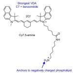 Development of a Highly Efficient Cancer Treatment Strategy using the Vibrionic-Driven Action of Synthetic Aminocyanines


Pingback: Molecular Scissors: An Introduction to Restriction Enzymes - Aneknowledge.com
Pingback: CRISPR Series: CRISPR and Gene Drive Systems: Advanced Mechanisms for Population Modification and Vector Control - Aneknowledge.com
Pingback: The Evolution of Plant Mutagenesis: Advanced Methodologies for Global Crop Improvement and Food Security - Aneknowledge.com