Cytoskeleton is a complex network of protein fibers and filaments that extends throughout the cytoplasm of a cell. Cellular processes such as crawling on substratum, cell division as well as muscle contraction is controlled by cytoskeleton. Three specific protein filaments make up the constituents of the cytoskeleton: Actin filaments (also called microfilaments), microtubules and intermediate filament. This article shall delve into the details regarding actin filaments and microtubules.
Actin filaments are the smallest elements of cytoskeleton ranging between 5-7nm in diameter.1 The length of the filament may vary greatly. The filaments are crosslinked into imperfect bundles or network. They are the most agile or flexible among the components of cytoskeleton. The actin filament is made of G-actin (globular actin) subunits. Actin is the most abundant and conserved protein present in eukaryotic cell.2 Actin filament is essential for various cellular processes including muscular contraction, maintenance of cell shape and cell locomotion. They are dynamic in nature and contain distinct polar ends. Microfilaments are constantly shrinking and growing in length, bundles and meshwork of microfilaments are continuously forming and dissolving.
Polymerization of Actin
Actin filament is synthesized by the polymerization of actin monomers (G-actin). The polymerization process of actin contains three main phases: nucleation phase, elongation phase and steady phase.
- Nucleation phase: In this phase, three G-actin come closer and bind together to form short unstable oligomers. When the oligomer reaches a certain length (generally three subunits) it acts as a stable nucleus.
- Elongation phase: The length of the filament increases by the addition of actin monomers to both ends of the filament. As length of the filament grows, the concentration of free G-actin decreases until an equilibrium is reached between filament and G-actin monomers.
- Steady phase: G-actin monomers is continuously exchanged with subunits at the filament ends but there is no net change in total length of the filament. The rate of association of actin subunit is equal to the rate of actin dissociation.

Myosin and Actin interaction
Myosin belong to a large superfamily of motor proteins. Its structure consists of three major parts: a motor domain, a coiled stalk and tail domain. The tail of the myosin molecule binds to the cargo (vesicle or other cellular processes) and carries it. The head of myosin is bound with an ATP molecule. It walks along the actin filament and the bound ATP is hydrolyzed during the process.
Different classes of myosin contain different functions. Myosin II found in the muscle cells form thick filaments which are used in muscular contraction. Myosin V has ability to take larger steps in the filament due to its longer neck region, making the transportation of cargo faster.2 Actin and myosin interaction is pivotal underlying event behind numerous cellular activities. Myosin head binds to specific sites on the actin filament initiating the actin-myosin interaction. The binding site is highly specific and regulated by various factors including the concentration of Calcium ions.

Role of Actin and Myosin in muscular contraction
When need for muscle contraction is perceived by the brain, it transmits electrical signals causing a cascade of reactions which ultimately leads to conformational change in tropomyosin. This leads to uncovering of binding sites for myosin on the actin filament. As the binding site gets exposed, the myosin head binds to the actin filament forming cross-bridges. The ATP attached to the myosin head is hydrolyzed generating mechanical energy required for the head to pivot. This action pulls the actin towards the center of the sarcomere, shortening the muscle fiber.2 Later in the cycle, ATP binds to myosin, causing its detachment from actin. This resets myosin for another round of contraction. Apart from muscular contraction, myosin and actin interaction is essential for a variety of cellular processes including endocytosis, cell division (particularly during cytokinesis), signal transduction, nutrient uptake, etc.
Function of active filament
- Muscle contraction: Actin interacts with myosin to generate force needed for muscular contraction.
- Structural support: Actin filament provides mechanical support to the cell and helps the organelles to assume their position in the cell.
- Intracellular transport: It coordinates the movement of various cell organelles, vesicle, and other cellular components within the cell.
- Cell division: It helps in the formation of mitotic spindle during cell division.
Microtubules are the largest among the cytoskeletal elements. They are filamentous structures composed of tubulin subunits. They are found scattered throughout the cytoplasm. They coordinate many essential movements in the cell which includes movement of cilia and flagella, intracellular transport of various cellular processes, segregation of chromosomes during cell division, etc.

Structure of Microtubule
Microtubule is made up of subunits called tubulin. Tubulin are heterodimers. They are composed of a-tubulin and b-tubulin subunits. a-tubulin and b-tubulin arrange themselves alternatively to form protofilament. Generally, 13 protofilament arrange themselves into a hollow cylindrical form to produce a microtubule. They are the largest among the cytoskeletal element found in the cell with the diameter about 25 nanometers.1 Due to their tube-like construction microtubules are much stiffer than microfilaments.2 Microtubule are dynamic structures. Like actin, microtubule also undergoes constant assembly and disassembly. Polarity is exhibited by microtubule filament. The microtubule filament consists of distinct + and – end.
Assembly and Disassembly of Microtubules
Cell contains two types of microtubules. One of them is stable and long-lived, another is unstable and has short life.2 Stable microtubule are found in non-replicating cell. They are present in some erythrocytes, platelets, cilia, flagella and neurons. Disassembly of these structures have catastrophic consequences: the sperms would be unable to swim, axons would retract and RBCs would lose their spring-like pliability.
Unstable short-lived microtubules are found in the cell which requires quick assembly and disassembly of microtubule structures.2 For instance, microtubules used in cell division has to quickly arrange themselves to form mitotic spindle and disassemble instantly in the interphase.
Microtubule assembly comprises of three major steps:
- a and b tubulins assemble to form protofilament.
- Protofilaments arrange themselves to form wall of microtubules.
- New tubulin subunits added to the end of the protofilaments elongates the microtubule.
GTP is bound to both a and b tubulins. Only the GTP bound to the b tubulin hydrolyzes to GDP. The binding affinity of tubulin for adjacent molecules decreases. Hence, disassembly of microtubules occur. Tubulin molecules bound to GDP are continuously lost from the – end and replaced by the addition of tubulin molecules bound to GTP on the + end.
Microtubules Organizing Center (MTOC)
MTOC is the structure found in eukaryotic cells from which microtubules emerge. It is essential for microtubules nucleation, stabilization and anchoring. The centrosome is an organelle that serves as the main MTOC of the animal cell as well as the regulator of cell cycle progression.1 MTOCS exists in basal bodies, centrioles at the pole of mitotic spindles in dividing cells, on chromosomes, etc. MTOCs have two major function:
- The organization of mitotic and meiotic spindle that separates the chromosomes during cell division.
- The organization of cilia and flagella.
Kinesin and Dynein
Kinesin and Dynein are microtubule associated motor proteins. Like any other motor proteins, kinesin and dynein use energy derived from hydrolysis of ATP to power wide variety of cellular functions. Dynein is involved in intracellular transport, cell division, beating movement of cilia and flagella. Kinesin also performs similar functions like arrangement of chromosomes in mitosis and meiosis as well as intracellular transport. Dynein shows a characteristic movement i.e. it moves toward the minus end of the microtubules. This movement is known as ‘retrograde transport’. The kinesins, however, moves towards the positive end of a microtubule. They transport the substances like protein and cellular components from the center of the cell towards the periphery. This movement is known as ‘anterograde movement’.
Function of cytoplasmic microtubules
- Mechanical function: The orientation of microtubules determines the shape of the cell, and cellular processes like axons, dendrites, microvilli, etc.
- Morphogenesis: Microtubules determines the shape of the developing cell during cell division.
- Contraction: The contraction of spindle fiber, movement of chromosomes, ciliary and flagellar motion depends upon microtubules.
- Intracellular transport: Microtubules are responsible for transport of macromolecule, granules and vesicle within the cell.1
REFERENCE
- Dr. P.S. Verma, Dr. C.K. Agrawal. Cell Biology, Genetics, Molecular Biology, Evolution and Ecology. fourteenth. (Bharatnagar S, Pradhan S, eds.). S.CHAND & COMPANY PVT.LTD.; 2016
- Lodish, Berk, Matsudaira, et al. Molecular Cell Biology. Fifth edition. W. H. Freeman; 2008.
Related posts:
 New Class of Antibiotics with a Novel Mode of Inhibition against World Health Organization’s Critical Listed Bacteria, Carbapenem-Resistant Acinetobacter baumannii
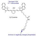 Development of a Highly Efficient Cancer Treatment Strategy using the Vibrionic-Driven Action of Synthetic Aminocyanines
 Natural Healers: Animals That Cure Themselves with Natural Solutions
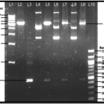 Factors affecting migration of Nucleic Acids in Agarose Gel Electrophoresis
 Scope and Applications of Bioinformatics