Majority of unicellular organisms and few multicellular organisms from identical copies of themselves through asexual reproduction. However, the multicellular and unicellular eukaryotes reproduce using sexual reproduction. In such higher organisms, specialized reproductive cells are produced known as gametes. The gametes produced by male are motile and are called sperm. The gametes produced by female are called ovum and they are non-motile. The male gamete fertilizes an ovum, forming a zygote. Zygote goes through a series of developmental changes until a new individual is formed. The somatic cells of the body that divides through mitosis contain diploid set of chromosomes. The gametes, however, are haploid. The fusion of two dissimilar haploid gamete results in formation of a diploid zygote. This haploidy is achieved by reduction division called meiosis.1 Meiosis divides the homologous pair of chromosomes into two sets so that each gamete obtains one of the sets. Meiocytes are those cells in which meiosis takes place.
1.1. Occurrence of Meiosis:
In animals, meiosis takes place just before the fertilization. In higher plants, meiosis and fertilization are separated by one or more cell generations. In lower plants, meiosis occurs before fertilization and the two events are separated by many cell generations. However, in unicellular organisms, meiosis may occur after fertilization.2
1.2. Stages of Meiosis:
Meiosis involves two consecutive cell divisions, which are; the first meiotic division and second meiotic division. Each division includes the following stages; prophase, metaphase, anaphase and telophase. The prophase of first meiotic division is long in which the homologous chromosomes become closely associated with each other and hereditary material is exchanged. The first meiotic division results in the formation of two haploid cells. The first meiotic division is division is also known as heterotypic division. In the second meiotic division, four haploid cells are created as the previously formed haploid cell divides mitotically. The second meiotic division is also referred to as the homotypic division.

The successive meiosis substages are briefly explained below:
- First Meiotic Division:
In G2 phase of interphase, a decisive change occurs that directs the cell towards meiosis instead of mitosis (Stern and Hotta,1968). At the beginning of first meiotic division, the nucleus of meiocyte starts to swell by absorption of water from the cytoplasm. Nuclear volume increase to three times. After these changes cell passes to the first stage of first meiotic division which is called prophase.
- Prophase I: It is the longest stage of the meiotic cell division and is much slower than the Prophase in mitosis. It includes the following substages:
i) Leptotene ii) Zygotene
iii) Pachytene iv) Diplotene
v) Diakinesis
i) Leptotene: The nuclear volume increases. Chromosomes appear like long, uncoiled and thin threads. The ends of the chromosomes converge towards the side of the nucleus where the centrosome lies. The centriole duplicates and each daughter centriole migrate towards the opposite poles of the cell. Each centriole divides on reaching at the poles.

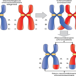
ii) Zygotene: The homologous chromosomes from father(sperm) and mother(ova) are attracted towards each other and their pairing takes place. The pairing of the homologous chromosomes is known as Synapsis and the homologous pair is called Bivalent. The synapsis can randomly occur at one or more points along the length of homologous chromosomes. The paired homologous chromosomes are joined by a proteinaceous framework called synaptonemal complex (SC). SC is involved in stabilizing the pairing of homologous chromosomes and facilitating crossing over.

iii) Pachytene: Nucleus grows in size. The bivalent pair splits up longitudinally to form a tetrad structure. Each chromatid in tetrad undergoes process of coiling and becomes shorter and thicker. Formation of Chiasma occurs and the two sister chromatids of each homologous pair of chromosome exchange segments. This phenomenon is called crossing over.
iv) Diplotene: The paired chromosomes begin to pull apart from each other but they do not separate completely as they are held together at the site of interchange called chiasmata (Singular: Chiasma). Often, unfolding of the chromatids take place, which allows for RNA synthesis and growth.
v) Diakinesis: The bivalent chromosomes become more condensed and they are distributed evenly in the nucleus. The nucleolus disappears and the nuclear envelope breaks down. The chiasma moves from the centromere towards the end of the chromosome and eventually the chiasmata diminish. The chromatids still remain connected to each other through the terminal chiasmata.
II) Metaphase I: Spindle fibres slowly attach themselves to the chromosomes and they fix the chromosomes in the equator region. Spindle fibres are attached to the centromere of homologous chromosomes through the involvement of their microtubules. Centromere of the chromosomes are directed towards opposite poles. There is increase in repulsive forces between the homologous chromosomes.
III) Anaphase I: Homologues are separated from each other. Each homologous chromosomes move towards the opposite poles of the cell. The chromosomes containing fewer chiasma tend to separate more rapidly than the one with many chiasmata. The two chromatids of chromosomes at opposite pole are not genetically identical due to crossing over (exchange of alleles in one of the chromatids)
IV) Telophase I: Haploid set of chromosomes arrives at each pole. Nuclei are reassembled. Endoplasmic reticulum forms the nuclear envelope around the chromosome and chromosomes uncoil. Then, the nucleolus reappears forming two daughter chromosomes. Cytokinesis occurs dividing the cell into two cells.
B) Second meiotic division: Division of each haploid meiocyte into two haploid cells occur in this phase. It includes 4 substages:
I) Prophase II: Each centriole divides into two and two pairs of centriole s are formed. Each pair migrates to the opposite pole. Spindle fibres are arranged. The nuclear membrane and nucleolus disappear. The chromosome becomes short and thick.
II) Metaphase II: The nuclear envelopes are completely broken down and the spindle fibre is fully formed. Each sister chromatid forms an individual kinetochore. The kinetochore attaches itself to microtubules from opposite sides. The sister chromatids are maximally condensed and aligned at the equator of the cell.1
III) Anaphase II: Chromosomal microtubules shorten. This moves the daughter chromosomes towards the opposite pole.
IV) Telophase II: The chromatids reach the opposite poles and then they are known as chromosomes. The endoplasmic reticulum forms nuclear envelope around the chromosome. The nucleolus reappears. After completion of karyokinesis, cytokinesis occurs in each haploid meiotic cells resulting in four haploid cells. All four cells have different types of chromosomes due to crossing over that occurred in Prophase I.3
1.3. Significance of Meiosis:
- A definite and constant number of the chromosome is maintained in the organism by meiosis division.
- Crossing over results in genetic variation. Variation is key to the progress in the evolutionary process.
1.4. Role of Meiosis in Genetic Variation:
Genetic variation is defined as the differences present in the genetic makeup of individuals belonging to same species. It is crucial for evolution. Favourable characteristics that arise trough variation are passed down to next generation for increasing their chances of survival. Meiosis contributes to genetic variation mainly through chromosomal rearrangement during crossing over and independent assortment of gametes. Meiosis ultimately forms four haploid cells that are neither identical to each other nor to their parent cell. The genetic information in the parent cell is distributed randomly i.e. independent assortment of gamete. The random assortment occurs during Metaphase I of meiosis I. During crossing over in Prophase I, the sites of interchange are chosen at random so they will be different in each cell gained through meiosis. This produces wide variety of recombinant chromosomes.
1.5. Diseases due to Meiosis dysfunction:
Different inherited disorders may occur if the chromosomes behave abnormally during meiosis. Chromosomal disorders may occur either due to abnormalities in chromosome number or chromosome structure rearrangement. Down Syndrome, Turner Syndrome, Jacobsen syndrome, certain cancers including chronic myelogenous leukaemia are result of abnormalities in chromosome number per cell. Chromosome structure may be rearranged by inversion or translocation. This may cause negative effects on development, fertility and even death.
1.6. Comparison between Meiosis and Mitosis
| Mitosis | Meiosis |
| I)Mitosis occurs continuously in the somatic cells. | Meiosis occurs in the germ cells (cells of testis or ovary) during the process of gametogenesis. |
| II)The whole process completes in one sequence or phase. | The whole process completes in two successive divisions that occurs one after another. |
| III) The prophase is of shorter duration and does not contain further substages. | The prophase is of longer duration and contains substages: Leptotene, Zygotene, Pachytene, Diplotene and Diakinesis |
| IV) No pairing or synapsis occurs between two homologous chromosomes. | Pairing or synapsis occurs between the two homologous chromosomes. |
| V) Crossing over does not take place. | Crossing over takes place. |
| VI) The telophase always occurs. | Telophase I is sometimes omitted. |
| VII) The chromosome number in the daughter cell remains same as the parent cell. | The chromosome number of daughter cell is reduced to half than the parent cells. |
| VIII) Two diploid cells are formed at the end of mitosis. | Four haploid cells are formed as a end product of meiosis. |
REFERENCE:
- Clark MA, Choi J, Douglas M. Biology 2e. Second. The Rice University Press; 2020.
- S.C. Rastogi. Cell and Molecular Biology. Third edition. New Age International Publishers
- Dr. P.S. Verma, Dr. C.K. Agrawal. Cell Biology, Genetics, Molecular Biology, Evolution and Ecology. fourteenth. (Bharatnagar S, Pradhan S, eds.). S.CHAND & COMPANY PVT.LTD.; 2016.
Related posts:
 Genomics: Types, Methods and Applications
 Comparison between Horizontal Gene Transfer Mechanisms in Bacteria: Transformation, Conjugation and Transduction
 Crossing Over: Mechanism, Types, Significance
 Mutation: Characteristics, Types and Applications
 DNA fingerprinting: History, Procedure and Its Applications